Paclitaxel-Conditioned Mesenchymal Stem Cells Enhance T Cell Activation and Cytotoxicity Against CD44+/CD24− Breast Cancer Stem Cells
DOI:
https://doi.org/10.48048/tis.2027.14177Keywords:
Breast cancer stem cells, Mesenchymal stem cells, Paclitaxel, T cell activation, Immunotherapy, CD44⁺/CD24⁻Abstract
Breast cancer stem cells (BCSCs) represent a critical population responsible for tumor recurrence, metastasis, and therapeutic resistance. Despite advances in immunotherapy, targeting BCSCs remains challenging due to their immune evasion mechanisms and limited T cell infiltration. To investigate the potential of paclitaxel-conditioned mesenchymal stem cells (MSCs) in enhancing T cell activation and cytotoxicity against CD44⁺/CD24⁻ breast cancer stem cells. BCSCs were isolated from MDA-MB-231 cells using magnetic-activated cell sorting (MACS) and characterized by flow cytometry. MSCs derived from umbilical cord blood were treated with paclitaxel (10 - 20 μg/mL) for 72 h to generate conditioned medium (CM-MSCs-Pax). CD8+ T cells were isolated from tumor-bearing BALB/c mice and activated using CM-MSCs-Pax. T cell activation was assessed by IL2RA (CD25) expression via qRT-PCR. Co-culture experiments evaluated cytotoxicity against BCSCs using the MTT assay, and molecular mechanisms were examined by analyzing caspase-3 and PD-L1 expression. MACS sorting successfully enriched CD44⁺/CD24⁻ BCSCs to 80.7% purity. Paclitaxel treatment significantly enhanced MSC secretion of IL-6, TNF-α, and IL-10 in conditioned medium. CM-MSCs-Pax treatment increased T cell activation, demonstrated by elevated IL2RA expression. Co-culture of activated T cells with BCSCs at a 10:1 ratio reduced BCSC viability to 9.68% at 72 h compared to 30.20% with unactivated T cells. This enhanced cytotoxicity was associated with increased caspase-3 expression and reduced PD-L1 expression in BCSCs. Paclitaxel-conditioned MSCs create a pro-inflammatory microenvironment that enhances T cell activation and cytotoxicity against BCSCs. This novel approach combining chemotherapy conditioning with immunotherapy represents a promising strategy for targeting therapy-resistant breast cancer stem cell populations.
HIGHLIGHTS
- Paclitaxel-conditioned MSCs (CM-MSCs-Pax) enhance CD8+ T cell activation against breast cancer stem cells (BCSCs).
- CM-MSCs-Pax increases pro-inflammatory cytokine secretion (IL-6, TNF-α, IL-10).
- Activated T cells exhibit significantly enhanced cytotoxicity against CD44+/CD24−
- BCSC viability decreases to ~9.7% at 72 h with CM-MSCs-Pax–activated T cells.
- Enhanced killing is associated with increased caspase-3 and reduced PD-L1 expression.
- Chemotherapy-conditioned MSCs offer a promising strategy to overcome BCSC immune resistance.
GRAPHICAL ABSTRACT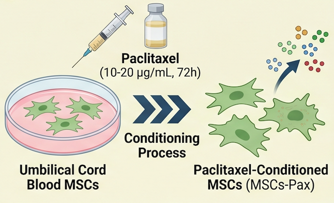
Downloads
References
SM Lima, RD Kehm and MB Terry. Global breast cancer incidence and mortality trends by region, age-groups, and fertility patterns. eClinicalMedicine 2021; 38, 100985.
H Zeng and G Lu. Research progress of breast cancer stem cell stemness and breast cancer recurrence. Journal of Biosciences and Medicines 2024; 12(8), 281-294.
Y Feng, M Spezia, S Huang, C Yuan, Z Zeng, L Zhang, X Ji, W Liu, B Huang, W Luo, B Liu, Y Lei, S Du, A Vuppalapati, HH Luu, RC Haydon, TC He and G Ren. Breast cancer development and progression: Risk factors, cancer stem cells, signaling pathways, genomics, and molecular pathogenesis. Genes & Diseases 2018; 5(2), 77-106.
W Shuo, Z Ying, C Weihong, L Jie, Z Yuren, F Huiting, X Yonggang, L Hongsheng and HS Lin. Breast cancer stem-like cells can promote metastasis by activating platelets and down-regulating antitumor activity of natural killer cells. Journal of Traditional Chinese Medicine 2016; 36(4), 530-537.
JS Crabtree and L Miele. Breast cancer stem cells. Biomedicines 2018; 6(3), 77.
D Hermansyah, S Syarifah, AM Muhar and A Putra. Unveiling paclitaxel-induced mesenchymal stem cells: Orchestrating Nrf2 modulation and apoptosis in CD44⁺/CD24⁻ cancer stem cells. Breast Cancer: Targets and Therapy 2024; 16, 319-328.
MA Elbaiomy, T Akl, N Atwan, AA Elsayed, M Elzaafarany and S Shamaa. Clinical impact of breast cancer stem cells in metastatic breast cancer patients. Journal of Oncology 2020; 2020, 2561726.
WC Sin and CL Lim. Breast cancer stem cells—from origins to targeted therapy. Stem Cell Investigation 2017; 4, 96.
LV Nalla, K Kalia and A Khairnar. Self-renewal signaling pathways in breast cancer stem cells. The International Journal of Biochemistry & Cell Biology 2019; 107, 140-153.
P Darvin, VS Nair and E Elkord. PD-L1 expression in human breast cancer stem cells is epigenetically regulated through posttranslational histone modifications. Journal of Oncology 2019; 2019, 3958908.
S Almozyan, D Colak, F Mansour, A Alaiya, O Al-Harazi, A Qattan, F Al-Mohanna, M Al-Alwan and H Ghebeh. PD-L1 promotes OCT4 and Nanog expression in breast cancer stem cells by sustaining PI3K/AKT pathway activation. International Journal of Cancer 2017; 141(7), 1402-1412.
J Chu, Y Li, X Fan, J Ma, J Li, G Lu, Y Zhang, Y Huang, W Li, X Huang, Z Fu, Y Yin and H Yuan. MiR-4319 suppress the malignancy of triple-negative breast cancer by regulating self-renewal and tumorigenesis of stem cells. Cellular Physiology and Biochemistry 2018; 48(2), 593-604.
T Li, C Zhang, Y Ding, W Zhai, K Liu, F Bu, T Tu, L Sun, W Zhu, F Zhou, W Qi, J Hu, H Chen and X Sun. Umbilical cord-derived mesenchymal stem cells promote proliferation and migration in MCF-7 and MDA-MB-231 breast cancer cells through activation of the ERK pathway. Oncology Reports 2015; 34(3), 1469-1477.
M Szewc, E Radzikowska-Bűchner, P Wdowiak, J Kozak, P Kuszta, E Niezabitowska, J Matysiak, K Kubiński and M Masłyk. MSCs as tumor-specific vectors for the delivery of anticancer agents—a potential therapeutic strategy in cancer diseases: Perspectives for quinazoline derivatives. International Journal of Molecular Sciences 2022; 23(5), 2745.
R Hass. Role of msc in the tumor microenvironment. Cancers 2020; 12(8), 2107.
HM Nawara, SM Afify, G Hassan, MH Zahra, A Seno and M Seno. Paclitaxel-based chemotherapy targeting cancer stem cells from mono- to combination therapy. Biomedicines 2021; 9(5), 500.
RC Sterner and RM Sterner. CAR-T cell therapy: Current limitations and potential strategies. Blood Cancer Journal 2021; 11(4), 69.
C D’Accardo, G Porcelli, LR Mangiapane, C Modica, VD Pantina, N Roozafzay, S Di Franco, M Gaggianesi, V Veschi, M Lo Iacono, M Todaro, A Turdo and G Stassi. Cancer cell targeting by CAR-T cells: A matter of stemness. Frontiers in Molecular Medicine 2022; 2, 1055028.
HE Marei, K Bedair, A Hasan, L Al-Mansoori, S Caratelli, G Sconocchia, A Gaiba and C Cenciarelli. Current status and innovative developments of CAR-T-cell therapy for the treatment of breast cancer. Cancer Cell International 2025; 25(1), 3.
H Wang, G Kaur, AI Sankin, F Chen, F Guan and X Zang. Immune checkpoint blockade and CAR-T cell therapy in hematologic malignancies. Journal of Hematology & Oncology 2019; 12(1), 59.
M Suzery, B Cahyono and ND Amalina. Citrus sinensis (L) peels extract inhibits metastasis of breast cancer cells by targeting the downregulation matrix metalloproteinases-9. Open Access Macedonian Journal of Medical Sciences 2021; 9(B), 464-469.
M Suzery, B Cahyono and ND Amalina. Antiproliferative and apoptosis effect of hyptolide from Hyptis pectinata (L.) Poit on human breast cancer cells. Journal of Applied Pharmaceutical Science 2020; 10(2), 001-006.
M Fredianto, H Herman, YD Ismiarto, A Putra, I Alif, ND Amalina and MA Nazar. Secretome of hypoxia-preconditioned mesenchymal stem cells enhance the expression of HIF-1a and bFGF in a rotator cuff tear model. Medicinski Glasnik 2023; 20(2), 242-248.
AM Muhar, F Makharim, D Hermansyah, A Putra, N Hidayah, ND Amalina and I Alif. Hypoxic mesenchymal stem cell-conditioned medium accelerates wound healing by regulating IL-10 and TGF-β levels in a full-thickness-wound rat model. Indonesian Journal of Biotechnology 2022; 27(4), 187-194.
YW Prajoko, A Putra, BT Dirja, AM Muhar and ND Amalina. The ameliorating effects of MSCs in controlling Treg-mediated B-cell depletion by indoleamine 2, 3-dioxygenase induction in PBMC of SLE patients. Open Access Macedonian Journal of Medical Sciences 2022; 10(A), 6-11.
BT Dirja, EH Wardoyo, A Putra and ND Amalina. Enhanced third degree burn wound healing by hypoxic mesenchymal stem cells’ secretome: IL-10 upregulation and TNF-α/PGE2 suppression. Trends in Sciences 2024; 21(10), 8288.
A Chaudhry, RM Samstein, P Treuting, Y Liang, MC Pils, JM Heinrich, RS Jack, FT Wunderlich, JC Brüning, W Müller and AY Rudensky. Interleukin-10 signaling in regulatory T cells is required for suppression of Th17 cell-mediated inflammation. Immunity 2011; 34(4), 566-578.
DA Paramita, D Hermansyah and ND Amalina. Regulation of p53 and survivin by Curcuma longa extract to caspase-3 dependent apoptosis in triple negative breast cancer cells. Medicinski Glasnik 2022; 19(2), 189-196.
Q Tang, Y Chen, X Li, S Long, Y Shi, Y Yu, W Wu, L Han and S Wang. The role of PD-1/PD-L1 and application of immune-checkpoint inhibitors in human cancers. Frontiers in Immunology 2022; 13, 964442.
MG Scioli, G Storti, F D’Amico, P Gentile, G Fabbri, V Cervelli and A Orlandi. The role of breast cancer stem cells as a prognostic marker and a target to improve the efficacy of breast cancer therapy. Cancers 2019; 11(7), 1021.
H Xu, Y Zhou, W Li, B Zhang, H Zhang, S Zhao, P Zheng, H Wu and J Yang. Tumor-derived mesenchymal-stem-cell-secreted IL-6 enhances resistance to cisplatin via the STAT3 pathway in breast cancer. Oncology Letters 2018; 15(6), 9142-9150.
DE Johnson, RA O'Keefe and JR Grandis. Targeting the IL-6/JAK/STAT3 signalling axis in cancer. Nature Reviews Clinical Oncology 2018; 15(4), 234-248.
T Hirano. IL-6 in inflammation, autoimmunity and cancer. International Immunology 2021; 33(3), 127-148.
H Jeong, J Koh, S Kim, J Yim, SG Song, H Kim, Y Li, SH Lee, YK Chung, H Kim, CH Lee, HY Kim, B Keam, SH Lee, DH Chung and YK Jeon. Cell-intrinsic PD-L1 signaling drives immunosuppression by myeloid-derived suppressor cells through IL-6/Jak/Stat3 in PD-L1-high lung cancer. Journal for ImmunoTherapy of Cancer 2025; 13(3), e010612.
SA Nish, D Schenten, T Wunderlich, SD Pope, Y Gao, N Hoshi, S Yu, X Yan, HK Lee, L Pasman, I Brodsky, B Yordy, H Zhao, J Bruning and R Medzhitov. T cell-intrinsic role of IL-6 signaling in primary and memory responses. eLife 2014; 3, e01949.
B Huang, X Lang and X Li. The role of IL-6/JAK2/STAT3 signaling pathway in cancers. Frontiers in Oncology 2022; 12, 1023177.
Y Wang, AHH Van Boxel-Dezaire, H Cheon, J Yang and GR Stark. STAT3 activation in response to IL-6 is prolonged by the binding of IL-6 receptor to EGF receptor. Proceedings of the National Academy of Sciences of the United States of America 2013; 110(42), 16975-16980.
FK Hartati, SB Widjanarko, TD Widyaningsih and M Rifa’i. Anti-inflammatory evaluation of black rice extract inhibits TNF-α, IFN-γ and IL-6 cytokines produced by immunocompetent cells. Food and Agricultural Immunology 2017; 28(6), 1116-1125.
A Putra, FB Ridwan, AI Putridewi, AR Kustiyah, K Wirastuti, NAC Sadyah, I Rosdiana and D Munir. The role of TNF-α induced MSCs on suppressive inflammation by increasing TGF-β and IL-10. Open Access Macedonian Journal of Medical Sciences 2018; 6(10), 1779-1783.
DM Darlan, D Munir, A Putra, I Alif, ND Amalina, NK Jusuf and IB Putra. Revealing the decrease of indoleamine 2,3-dioxygenase as a major constituent for B cells survival post-mesenchymal stem cells co-cultured with peripheral blood mononuclear cell (PBMC) of systemic lupus erythematosus (SLE) patients. Medicinski Glasnik 2022; 19(1), 12-18.
J Xi, M Xu, Z Song, H Li, S Xu, C Wang, H Song and J Bai. Stimulatory role of interleukin 10 in CD8+ T cells through STATs in gastric cancer. Tumor Biology 2017; 39(5), 1010428317706209.
Z Guo, Q He, Y Zhang, Y Li and Z Zhang. Mechanisms of interleukin-10-mediated immunosuppression in viral infections. Pathogens 2025; 14(10), 989.
G Wang, D Zhang, S Yang, Y Wang, Z Tang and X Fu. Co-administration of genistein with doxorubicin-loaded polypeptide nanoparticles weakens the metastasis of malignant prostate cancer by amplifying oxidative damage. Biomaterials Science 2018; 6(4), 827-835.
ND Amalina, IP Nurhayati and E Meiyanto. Doxorubicin induces lamellipodia formation and cell migration. Indonesian Journal of Cancer Chemoprevention 2017; 8(2), 61-67.
S Ran. The role of TLR4 in chemotherapy-driven metastasis. Cancer Research 2015; 75(12), 2405-2410.
J Abdi, I Rashedi and A Keating. Concise review: TLR pathway-miRNA interplay in mesenchymal stromal cells: Regulatory roles and therapeutic directions. Stem Cells 2018; 36(11), 1655-1662.
LW Pfannenstiel, SS Lam, LA Emens, EM Jaffee and TD Armstrong. Paclitaxel enhances early dendritic cell maturation and function through TLR4 signaling in mice. Cellular Immunology 2010; 263(1), 79-87.
MA Huseni, L Wang, JE Klementowicz, K Yuen, B Breart, C Orr, LF Liu, Y Li, V Gupta, C Li, D Rishipathak, J Peng, Y Şenbabaoǧlu, Z Modrusan, S Keerthivasan, S Madireddi, YJ Chen, EJ Fraser, N Leng, H Hamidi, H Koeppen, J Ziai, K Hashimoto, M Fassò, P Williams, DF McDermott, JE Rosenberg, T Powles, LA Emens, PS Hegde, I Mellman, SJ Turley, MS Wilson, S Mariathasan, L Molinero, M Merchant and NR West. CD8+ T cell-intrinsic IL-6 signaling promotes resistance to anti-PD-L1 immunotherapy. Cell Reports Medicine 2023; 4(1), 100878.
X Pu, SJ Storr, Y Zhang, EA Rakha, AR Green, IO Ellis and SG Martin. Caspase-3 and caspase-8 expression in breast cancer: Caspase-3 is associated with survival. Apoptosis 2017; 22(3), 357-368.
KM Zak, P Grudnik, K Magiera, A Dömling, G Dubin and TA Holak. Structural biology of the immune checkpoint receptor PD-1 and its ligands PD-L1/PD-L2. Structure 2017; 25(8), 1163-1174.
M Elfoly, JY Mirza, A Alaiya, AA Al-Hazzani, A Tulbah, M Al-Alwan and H Ghebeh. PD-L1 intrinsically promotes the proliferation of breast cancer cells through the SKP2-p27/p21 axis. Cancer Cell International 2024; 24(1), 161.
M Jin, J Fang, J Peng, X Wang, P Xing, K Jia, J Hu, D Wang, Y Ding, X Wang, W Li and Z Chen. PD-1/PD-L1 immune checkpoint blockade in breast cancer: Research insights and sensitization strategies. Molecular Cancer 2024; 23(1), 266.
Downloads
Published
How to Cite
Issue
Section
License
Copyright (c) 2026 Walailak University

This work is licensed under a Creative Commons Attribution-NonCommercial-NoDerivatives 4.0 International License.