
Therapeutic Potential of Passiflora edulis Sims Seed Extract on Molecular and Histological Markers of Wound Healing in a Diabetic Rat Model
DOI:
https://doi.org/10.48048/tis.2026.11755Keywords:
Passiflora edulis sims, Passiflora edulis sims seed extract (PFSE), Diabetes mellitus, Wound healing, Oxidative stress (MDA), Inflammation (TNF-α), Apoptosis (Caspase-3), Epidermal growth factor (EGF), Collagen deposition, EpithelializationAbstract
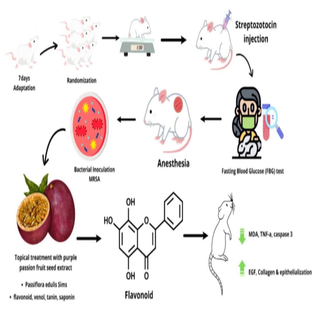
Diabetes mellitus is a chronic metabolic disorder characterized by persistent hyperglycemia and oxidative stress, which contribute to vascular dysfunction, tissue damage, hypoxia, and an increased risk of foot ulceration and amputation. Uncontrolled hyperglycemia elevates reactive oxygen species (ROS), increases malondialdehyde (MDA) production, and activates the NF-κB signaling pathway, leading to the release of pro-inflammatory cytokines such as TNF-α, IL-1, and IL-6. This cascade promotes caspase-3 activation and inflammatory cell damage in keratinocytes and fibroblasts, impairing extracellular matrix formation and re-epithelialization. Although antioxidant defenses play a critical role in wound healing, scientific validation of natural topical agents remains limited. Seeds of Passiflora edulis Sims, rich in flavonoids, gallic acid derivatives, and antioxidant compounds with anti-inflammatory and antimicrobial properties, have been identified as promising candidates for wound therapy. This study investigated molecular pathways involving epidermal growth factor (EGF), MDA, TNF-α, collagen deposition, and epithelialization as key regulators of oxidative stress, inflammation, and tissue repair. A post-test-only experimental design was conducted using 25 male Wistar rats with streptozotocin-nicotinamide-induced diabetes, divided into 5 groups: Negative control (ointment base), positive control (Tribee salf), and 3 treatment groups receiving Passiflora edulis seed extract (PFSE) at doses of 50, 100, and 150 mg/g. Molecular markers were analyzed using ELISA (MDA, EGF, caspase-3), immunohistochemistry (TNF-α), and histological staining (Masson’s Trichrome for collagen and hematoxylin-eosin for epithelialization). Data were statistically evaluated using ANOVA with Bonferroni post hoc, independent t-tests, and Kruskal-Wallis tests, with significance set at p < 0.05. The results demonstrated that PFSE significantly reduced MDA, TNF-α, and caspase-3 levels, while increasing EGF expression, collagen deposition, and epithelialization compared with the control groups (p < 0.05). These findings highlight the therapeutic potential of Passiflora edulis seed extract as an antioxidant, anti-inflammatory, and wound-healing agent for diabetic wounds.
HIGHLIGHTS
Experimental Model
- 25 male Wistar rats, adapted for 7 days.
- 5 groups: Negative control (ointment base), positive control (Tribee salf), and 3 treatment groups with Passiflora edulis Sims seed extract (PFSE 50, 100 and 150 mg/g).
- Diabetes was induced using streptozotocin (45 mg/kg, i.p.) combined with nicotinamide (110 mg/kg, i.p.).
- Wounds were created by punch biopsy followed by MRSA inoculation (10⁶ CFU).
- Treatment: Topical application of PFSE according to dosage.
Assessment Parameters
- Biomarkers: MDA, Caspase-3, and EGF (ELISA); TNF-α (immunohistochemistry).
- Histology: Collagen (Masson’s Trichrome) and epithelialization (H&E).
- Key findings: PFSE accelerated wound healing in diabetic rats infected with MRSA by modulating oxidative stress, inflammation, apoptosis, and tissue repair biomarkers.
GRAPHICAL ABSTRACT
Downloads
References
S Yang, Y Li, C Liu, Y Wu, Z Wan and D Shen. Pathogenesis and treatment of wound healing in patients with diabetes after tooth extraction. Frontiers in Endocrinology 2022; 13, 949535.
A Chaudhary, S Bag, P Banerjee and J Chatterjee. Wound healing efficacy of Jamun honey in diabetic mice model through reepithelialization, collagen deposition and angiogenesis. Journal of Traditional and Complementary Medicine 2020; 10(6), 529-543.
V Mujica, R Orrego, R Fuentealba, E Leiva and J Zúñiga-Hernández. Propolis as an adjuvant in the healing of human diabetic foot wounds receiving care in the diagnostic and treatment centre from the regional hospital of Talca. Journal of Diabetes Research 2019; 2019(1), 2507578.
Y Zheng, SH Ley and FB Hu. Global aetiology and epidemiology of type 2 diabetes mellitus and its complications. Nature Reviews Endocrinology 2018; 14(2), 88-98.
M Fazil and S Nikhat. Topical medicines for wound healing: A systematic review of Unani literature with recent advances. Journal of Ethnopharmacology 2020; 257, 112878.
J Holl, C Kowalewski, Z Zimek, P Fiedor, A Kaminski, T Oldak, M Moniuszko, A Eljaszewicz and S Steiger. Cells chronic diabetic wounds and their treatment with skin substitutes. Cells 2021; 10(3), 655.
T Tomita. Apoptosis in pancreatic β-islet cells in Type 2 diabetes. Bosnian Journal of Basic Medical Sciences 2016; 16(3), 162-179.
NX Landén, D Li and M Ståhle. Transition from inflammation to proliferation: A critical step during wound healing. Cellular and Molecular Life Sciences 2016; 73(20), 3861-3885.
M Zhra, RJ Qasem, F Aldossari, R Saleem and A Aljada. A comprehensive exploration of caspase detection methods: From classical approaches to cutting-edge innovations. International Journal of Molecular Sciences 2024; 25(10), 5460.
P Luka, A Cellular, D Biomolecular and Y Moenadjat. Wound healing: Cellular and biomolecular aspects. Universitas Indonesia, Jawa Barat, Indonesia, 2023.
L Cañedo-Dorantes and M Cañedo-Ayala. Skin acute wound healing: A comprehensive review. International Journal of Inflammation 2019; 2019(1), 3706315.
L Moretti, J Stalfort, TH Barker and D Abebayehu. The interplay of fibroblasts, the extracellular matrix, and inflammation in scar formation. Journal of Biological Chemistry 2022; 298(2), 101530.
M Kumar. Different blood collection methods from rats: A review. Balneo Research Journal 2017; 8(2), 46-50.
EI Morgun and EA Vorotelyak. Epidermal stem cells in hair follicle cycling and skin regeneration: A view from the perspective of inflammation. Frontiers in Cell and Developmental Biology 2020; 8, 581697.
VP Nakhate, NS Akojwar, SK Sinha, AD Lomte, M Dhobi, PR Itankar and SK Prasad. Wound healing potential of Acacia catechu in streptozotocin-induced diabetic mice using in vivo and in silico approach. Journal of Traditional and Complementary Medicine 2023; 13(5), 489-499.
C Loretelli, M Ben Nasr, G Giatsidis, R Bassi, L Lancerotto, F D’Addio, A Valderrama-Vasquez, S Scherer, L Salvatore, M Madaghiele, A Abdelsalam, E Ippolito, E Assi, V Usuelli, B El Essawy, A Sannino, G Pietramaggiori, GV Zuccotti, DP Orgill and P Fiorina. Embryonic stem cell extracts improve wound healing in diabetic mice. Acta Diabetologica 2020; 57(7), 883-890.
Z Jafari, H Bardania, MJ Barmak, S Eslami, Y Mahmoudi-Mourderaz, N Roustaei, MS Talebianpoor, EP Kokhdan and SS Khoramrooz. Antimicrobial, anti-inflammatory, and wound healing properties of myrtus communis leaf methanolic extract ointment on burn wound infection induced by methicillin-resistant Staphylococcus aureus in rats. BioMed Research International 2024; 2024(1), 6758817.
SE Davis, SS Tulandi, OS Datu, F Sangande and DN Pareta. Formulation and evaluation of ointment preparations of ethanol extract of Hibiscus rosa-sinensis L. leaves with various ointment bases. Jurnal Biofarmasetikal Tropis 2022; 10(2), 86-88.
S Güzel, Y Özay, M Kumaş, C Uzun, EG Özkorkmaz, Z Yıldırım, M Ülger, G Güler, A Çelik, Y Çamlıca and A Kahraman. Wound healing properties, antimicrobial and antioxidant activities of Salvia kronenburgii Rech. f. and Salvia euphratica Montbret, Aucher & Rech. f. var. euphratica on excision and incision wound models in diabetic rats. Biomedicine and Pharmacotherapy 2019; 111, 1260-1276.
M Zamanifard, M Nasiri, F Yarahmadi, S Zonoori, O Razani, Z Salajegheh, M Imanipour, SM Mohammadi, N Jomehzadeh and M Asadi. Healing of diabetic foot ulcer with topical and oral administrations of herbal products: A systematic review and meta-analysis of randomized controlled trials. International Wound Journal 2024; 21(2), e14760.
NK Jusuf, IB Putra and NK Dewi. Antibacterial activity of passion fruit purple variant (Passiflora edulis sims var. edulis) seeds extract against propionibacterium acnes. Clinical, Cosmetic and Investigational Dermatology 2020; 13, 99-104.
BM Bakadia, AAQ Ahmed, L Lamboni, Z Shi, BM Mukole, R Zheng, MP Mbang, B Zhang, M Gauthier and G Yang. Engineering homologous platelet-rich plasma, platelet-rich plasma-derived exosomes, and mesenchymal stem cell-derived exosomes-based dual-crosslinked hydrogels as bioactive diabetic wound dressings. Bioactive Materials 2023; 28, 74-94.
Bureau of Central Statistics (BCS). Indonesian seasonal fruit and vegetable crop statistics. Directorate General of Horticulture, Central Bureau of Statistics, Jakarta, Indonesia, 2020.
AE Marpaung, N Karsinah and BB Karo. Characterization and evaluation of passion fruit acid hybrid from purple and red passion fruit acid crossing. Jurnal Hortikultura 2015; 26(2), 163-170.
W Sukketsiri, S Daodee, S Parhira, W Malakul, S Tunsophon, N Sutthiwong, S Tanasawet and P Chonpathompikunlert. Chemical characterization of Passiflora edulis extracts and their in vitro antioxidant, anti-inflammatory, anti-lipid activities, and ex-vivo vasodilation effect. Journal of King Saud University - Science 2023; 35(1), 102431.
F Brahmi, I Mateos-Aparicio, K Mouhoubi, S Guemouni, T Sahki, F Dahmoune, F Belmehdi, C Bessai, K Madani and L Boulekbache-Makhlouf. Kinetic modeling of convective and microwave drying of potato peels and their effects on antioxidant content and capacity. Antioxidants 2023; 12(3), 638.
J Lang, SE Ramos, M Smohunova, L Bigler and MC Schuman. Screening of leaf extraction and storage conditions for eco-metabolomics studies. Plant Direct 2024; 8(4), e578
V Handayani, MAN Fauzan, AR Nadya, H Widiastuti, A Malik and AR Ahmad. Optimization of the microwave-assisted extraction method of passion fruit seeds (Passiflora edulis Sims) on antioxidant activity. JFIOnline 2025; 17(1), 9-14.
M Muslim, NK Jusuf and IB Putra. The effect of 3% passion fruit purple variant (Passiflora edulis Sims var. Edulis) seed extract cream on facial skin aging. Journal of Pakistan Association of Dermatologists 2023; 33(2), 566-573.
N Sharma. Comparison of wound healing properties of herbal ointments with povidone iodine on basis of histological changes. Journal of Animal Research 2019; 9(1), 173-177.
N Sharma, R Singh, S Jawre, R Vaish and MKP Chauhan. Pharmacological and therapeutic properties of Jasminum officinale. L: A review. Indian Journal of Ecology 2022; 49(3), 1122-1128.
R Tungadi and MS Pakaya. Formulasi dan evaluasi stabilitas fisik sediaan krim senyawa astaxanthin (in Indonesian). Indonesian Journal of Pharmaceutical Education 2023; 3(1), 117-124.
ED Sersemova, M Arev, P Apostolova, D Karpicarov, V Maksimova, D Miceva, A Cvetkovski and M Samardziska. Preparation and characterization of amphiphilic cream formulations with meloxicam. Pharmacia 2024; 71, 1-7.
Y Abhishek and S Krishanu. Formulation and evaluation of herbal ointment using Emblica officinalis extract. World Journal of Advanced Research and Reviews 2021; 9(2), 032-037.
Q Zhao, J Xu, X Han, Z Zhang, J Qu and Z Cheng. Growth differentiation factor 10 induces angiogenesis to promote wound healing in rats with diabetic foot ulcers by activating TGF-β1/Smad3 signaling pathway. Frontiers in Endocrinology 2023; 13, 1013018.
G Azkona. Implementing the 3Rs in laboratory animal research - from theory to practice. Animals 2023; 13(19), 3063.
KY Cheng, ZH Lin, YP Cheng, HY Chiu, NL Yeh, TK Wu and JS Wu. Wound Healing in Streptozotocin-Induced Diabetic Rats Using Atmospheric-Pressure Argon Plasma Jet. Scientific Reports 2018; 8(1), 12214.
DS Masson-Meyers, TAM Andrade, GF Caetano, FR Guimaraes, MN Leite, SN Leite and MAC Frade. Experimental models and methods for cutaneous wound healing assessment. International Journal of Experimental Pathology 2020; 101(1-2), 21-37.
MB Rowland, PE Moore, C Bui and RN Correll. Assessing wound closure in mice using skin-punch biopsy. STAR Protocols 2023; 4(1), 101989.
AB Hora, LS Biano, ACS Nascimento, ZT Camargo, GI Heiden, RLC Albulquerque-Júnior, R Grespan, JMD Aragão and EA Camargo. Isoorientin improves excisional skin wound healing in mice. Pharmaceuticals 2024; 17(10), 1368.
MS Rahman, N Takahashi, T Iwabuchi, T Nishimura, T Harada, A Okumura, N Takei, Y Nomura and KJ Tsuchiya. Elevated risk of attention deficit hyperactivity disorder (ADHD) in Japanese children with higher genetic susceptibility to ADHD with a birth weight under 2000 g. BMC Medicine 2021; 19(1), 229.
A Herman and AP Herman. Herbal products and their active constituents for diabetic wound healing - preclinical and clinical studies: A systematic review. Pharmaceutics 2023; 15(1), 281.
A Fauzi, D Vidiastuti, A Noviatri and MY Rizal. Blueberry extract (Vaccinium corymbosum) attenuates Tnf-α expression and renal inflammatory cell counts in rats models of acute kidney injury. Advances in Animal and Veterinary Sciences 2021; 9(7), 1087-1094.
K Sharun, SA Banu, M Mamachan, A Subash, K Mathesh, R Kumar, OR Vinodhkumar, K Dhama, L Abualigah, AM Pawde and Amarpal. Comparative evaluation of masson’s trichrome and picrosirius red staining for digital collagen quantification using ImageJ in rabbit wound healing research. Journal of Experimental Biology and Agricultural Sciences 2023; 11(5), 822-833.
Frieda, I Julianto, N Dharmawan, A Kusumawardani, N Adi and EY Ellistasari. Description of type I collagen deposition according to age in Wistar strain rats: An in vivo study. Medika Kartika: Jurnal Kedokteran dan Kesehatan 2022; 5(2), 183-194.
M Van De Vyver, K Boodhoo, T Frazier, K Hamel, M Kopcewicz, B Levi, M Maartens, S Machcinska, J Nunez, C Pagani, E Rogers, K Walendzik, J Wisniewska, B Gawronska-Kozak and JM Gimble. Histology scoring system for murine cutaneous wounds. Stem Cells and Development 2021; 30(23), 1141-1152.
B Srinivasan and MD Lloyd. Dose-response curves and the determination of IC50 and EC50 values. Journal of Medicinal Chemistry 2024; 67(20), 17931-1793.
K Abbasi, S Tavakolizadeh, A Hadi, M Hosseini, RS Soufdoost, A Heboyan, M Alam and S Fani-Hanifeh. The wound healing effect of collagen/adipose-derived stem cells (ADSCs) hydrogel: In vivo study. Veterinary Medicine and Science 2023; 9(1), 282-289.
Y Sharma, A Kaur, R Bhardwaj, N Srivastava, M Lal, S Madan and K Bala. Preclinical assessment of stem of Nicotiana tabacum on excision wound model. Bioorganic Chemistry 2021; 109, 104731.
M Min, C Egli, RA Bartolome and RK Sivamani. Ex vivo evaluation of a liposome-mediated antioxidant delivery system on markers of skin photoaging and skin penetration. Clinical, Cosmetic and Investigational Dermatology 2024; 17, 1481-1494.
RNF Genuino, BL Dofitas, MCFR Batac, MBTG Pascual and AA Abrilla. Systematic review and meta-analysis on synthetic antifungal versus keratolytic agents for topical treatment of pityriasis versicolor. Acta Medica Philippina 2024; 58(1), 64-78.
NR Hartaman, Z Abidin and A Dahlia. Antioxidant activity of porang tuber (Amorphophallus oncophyllus) ethanol extract using ultrasonic extraction method. Makassar Natural Product Journal 2023; 1(3), 2023-2155.
S Aydin, E Emre, K Ugur, MA Aydin, İ Sahin, V Cinar and T Akbulut. An overview of ELISA: A review and update on best laboratory practices for quantifying peptides and proteins in biological fluids. Journal of International Medical Research 2025; 53(2), 03000605251315913.
RA Karas, S Alexeree, H Elsayed and YA Attia. Assessment of wound healing activity in diabetic mice treated with a novel therapeutic combination of selenium nanoparticles and platelets rich plasma. Scientific Reports 2024; 14(1), 5346.
S Polaka, P Katare, B Pawar, N Vasdev, T Gupta, K Rajpoot, P Sengupta and RK Tekade. Emerging ROS modulating technologies for augmentation of the wound healing process. ACS Omega 2022; 7(35), 30657-30672.
M Siniawska and A Wojdyło. Polyphenol profiling by LC QTOF/ESI-MS and biological activity of purple passion fruit epicarp extract. Molecules 2023; 28(18), 6711.
AY Wirya, KK Winaya, IGN Darmaputra, N Suryawati, IGAAD Karmila and NMD Puspawati. Topical application of purple sweet potato (Ipomoea batatas L.) extract cream increases superoxide dismutase (SOD) levels and decreases caspase-3 levels in rats (Rattus norvegicus) with photoaging due to ultraviolet B exposure. Intisari Sains Medis 2023; 14(2), 544-551.
SA Tajammal, A Coffey and SP Tan. Green tea polyphenols in wound healing: Therapeutic mechanisms, potential applications and challenges in commercial use for diabetic wound healing. Processes 2025; 13(3), 653.
J Berlanga-Acosta, A Garcia-Ojalvo, J Fernández-Montequin, V Falcon-Cama, N Acosta-Rivero, G Guillen-Nieto, M Pujol-Ferrer, M Limonta-Fernandez, M Ayala-Avila and E Eriksson. Epidermal growth factor intralesional delivery in chronic wounds: The pioneer and standalone technique for reversing wound chronicity and promoting sustainable healing. International Journal of Molecular Sciences 2024; 25(20), 10883.
DM Afrida. 2018, The effect of oral and topical administration of sweet orange (Citrus sinensis) peel extract on TNF-α expression and collagen density in the wound healing process of incision in diabetic mellitus rats. Undergraduate Thesis. Universitas Brawijaya, Jawa Timur, Indonesia.
AJ Didier, J Stiene, L Fang, D Watkins, LD Dworkin and JF Creeden. Antioxidant and anti-tumor effects of dietary vitamins A, C, and E. Antioxidants 2023; 12(3), 632.
DVD Vlekkert, E Machado and A d'Azzo. Analysis of Generalized Fibrosis in Mouse Tissue Sections with Masson's Trichrome Staining. Bio-protocol 2020; 10(10), e3629
N Dasari, A Jiang, A Skochdopole, J Chung, EM Reece, J Vorstenbosch and S Winocour. Updates in diabetic wound healing, inflammation, and scarring. Seminars in Plastic Surgery 2021; 35(3), 153-158.
R Sklenářová, N Akla, MJ Latorre, J Ulrichová and J Franková. Collagen as a biomaterial for skin and corneal wound healing. Journal of Functional Biomaterials 2022; 13(4), 249.
J Song, K Zhu, H Wang, M Wu, Y Wu and Q Zhang. Deciphering the emerging role of programmed cell death in diabetic wound healing. International Journal of Biological Sciences 2023; 19(15), 4989-5003.
G Weyya, A Belay and E Tadesse. Passion fruit (Passiflora edulis Sims) by-products as a source of bioactive compounds for non-communicable disease prevention: Extraction methods and mechanisms of action: A systematic review. Frontiers in Nutrition 2024; 11, 1340511.
Published
How to Cite
Issue
Section
License
Copyright (c) 2025 Walailak University

This work is licensed under a Creative Commons Attribution-NonCommercial-NoDerivatives 4.0 International License.


