A Comparative Evaluation of the Vasorelaxant Effects of F-1 and F-3 on Aorta and Pulmonary artery: Integrated In Vitro, In Vivo and In Silico Approaches
DOI:
https://doi.org/10.48048/tis.2027.14004Keywords:
F-1 compound, F-3 compound, Vasorelaxation, Vascular smooth muscle, Aorta, Pulmonary artery, Molecular dockingAbstract
This study aimed to evaluate the vasomodulatory and antihypertensive effects of compounds 1-Phenyl-6,7-dimethoxy-1,2,3,4-tetrahydroisoquinoline hydrochloride (F-1) and 1-(3′-hydroxyphenyl)-6,7-dimethoxy-1,2,3,4-tetrahydroisoquinoline hydrochloride (F-3) using an integrated approach combining in silico molecular docking, in vitro functional assays, and in vivo experimental models. Molecular docking analysis revealed that both compounds exhibit high binding affinity toward calcium-regulating targets, particularly the L-type calcium channel (Cav1.2), with binding energies of −8.2 kcal/mol (F1) and −8.1 kcal/mol (F3), indicating stable ligand–protein interactions and potential modulation of calcium influx. In vitro experiments demonstrated that both compounds significantly inhibited phenylephrine-induced vascular smooth muscle contraction in a concentration-dependent manner. F1 exhibited higher potency (IC₅₀ = 3.1×10⁻⁷ M) compared to F3 (IC₅₀ = 4.29×10⁻⁶ M), showing a more consistent and gradual inhibitory profile. Additionally, both compounds enhanced the vasorelaxant effect of verapamil and partially attenuated the inhibitory action of phentolamine, suggesting involvement in both calcium channel modulation and adrenergic signaling pathways. The reduction of their effects in the presence of L-NAME indicates partial dependence on nitric oxide-mediated mechanisms. In vivo studies in adrenaline-induced hypertensive rats demonstrated that F1 and F3 significantly reduced systolic and diastolic blood pressure in a dose- and time-dependent manner at doses of 10, 25, and 50 mg/kg. F1 showed a more pronounced antihypertensive effect, effectively restoring blood pressure toward physiological levels. Overall, the findings indicate that F1 is a more potent vasorelaxant and antihypertensive agent than F3. The observed effects are mediated through a multi-target mechanism involving inhibition of calcium influx, modulation of adrenergic signaling, and partial involvement of nitric oxide pathways. These results highlight F1 as a promising candidate for the development of novel therapeutic agents for the treatment of hypertension and vascular dysfunction.
HIGHLIGHTS
- F1 and F3 demonstrated significant vasorelaxant activity in vitro and ex vivo models
- Both compounds inhibited KCl- and phenylephrine-induced vascular contractions in a concentration-dependent manner
- F1 exhibited higher potency and efficacy compared to F3 across all experimental conditions
- Vasorelaxant effects were partially independent of nitric oxide, as confirmed using L-NAME
- In vivo studies showed marked reductions in systolic and diastolic blood pressure
- F1 produced stronger and more sustained antihypertensive effects than F3
- Molecular docking revealed high binding affinity to L-type (Cav1.2) and R-type calcium channels
- Interactions were mainly mediated by hydrophobic and π-related mechanisms
- Calcium channel modulation is a key mechanism underlying the observed pharmacological effects
- F1 is a promising candidate for further development as a cardiovascular therapeutic agent
GRAPHICAL ABSTRACT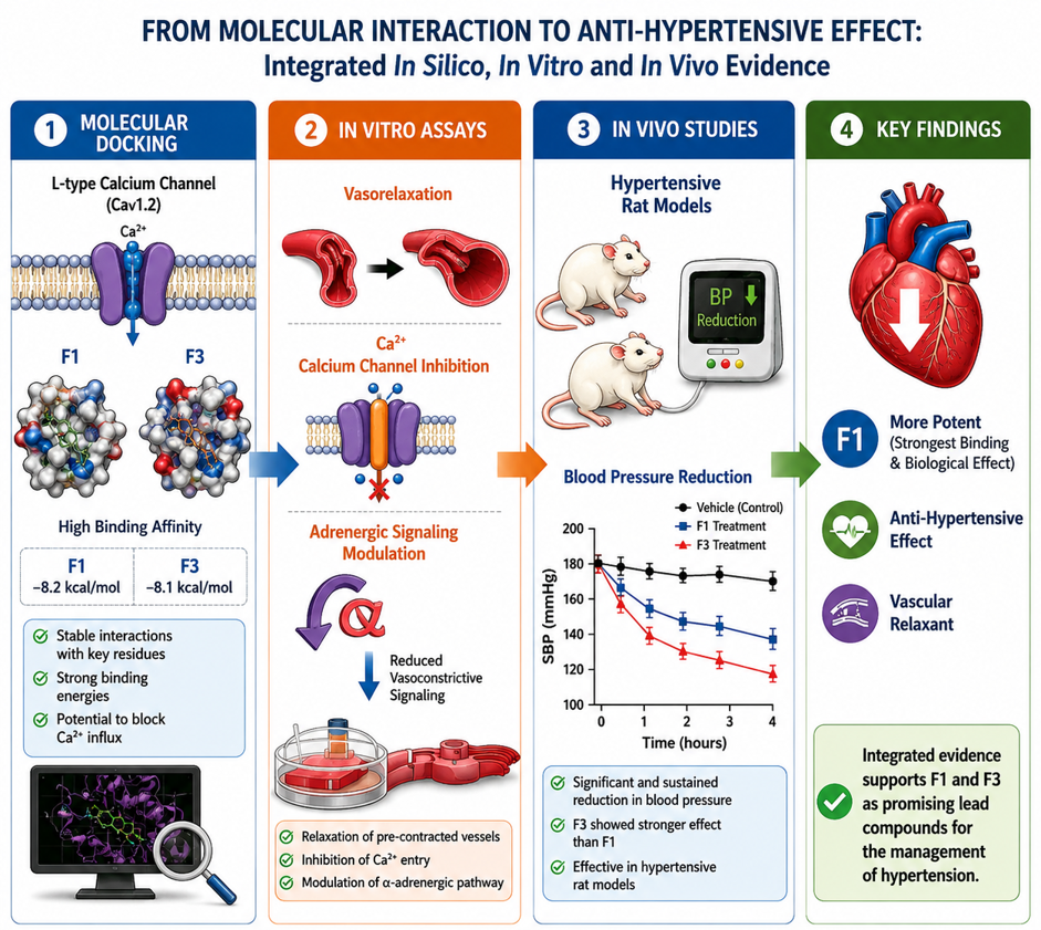
Downloads
References
MSh Bakieva, SR Rustamova, TO Rakhmonov, NN Sharipova and KS Mukhitdinova. Hypotensive effect of the alkaloid benzoylheteratisine on the functional activity of rat aortic smooth muscle cells. Academic Research Journal 2022; 1(2), 24-30.
J Park, S Shin, Y Kim, Y Bu, HY Choi and K Lee. Vasorelaxant and antihypertensive effects of the Cibotii Rhizoma hydroethanolic extract via the PI3K/AKT/eNOS pathway. Phytomedicine 2025; 148, 157437.
KKY Isla, MM Tanae, MTR de Lima-Landman, PM de Magalhães, AJ Lapa and C Souccar. Vasorelaxant effects of ellagitannins isolated from Cuphea carthagenensis. Planta Medica 2024; 90(4), 276-285.
C Chen, C Guo, J Gao, K Shi, J Cheng, J Zhang, S Chen, Y Liu and A Liu. Vasorelaxant and antihypertensive effects of Tianshu Capsule on rats: An in vitro and in vivo approach. Biomedicine & Pharmacotherapy 2019; 111, 188-197.
J Jung, S Shin, J Park, K Lee and HY Choi. Hypotensive and vasorelaxant effects of Sanguisorbae Radix ethanol extract in spontaneously hypertensive and Sprague Dawley rats. Nutrients 2023; 15(21), 4510.
P Wicha, A Onsa-Ard, W Chaichompoo, A Suksamrarn and C Tocharus. Vasorelaxant and antihypertensive effects of neferine in rats: An in vitro and in vivo study. Planta Medica 2020; 86(7), 496-504.
N Shobako, A Ishikado, Y Ogawa, Y Sono, T Kusakari, M Suwa, M Matsumoto and K Ohinata. Vasorelaxant and antihypertensive effects that are dependent on the endothelial NO system exhibited by rice bran-derived tripeptide. Journal of Agricultural and Food Chemistry 2019; 67(5), 1437-1442.
S Amtaghri, A Amssayef, M Slaoui and M Eddouks. Antihypertensive and vasorelaxant effects of Hibiscus rosa-sinensis through angiotensin-converting enzyme-2 (ACE-2), and Ca2+ channels pathways. Cardiovascular & Hematological Disorders-Drug Targets 2022; 22(1), 27-37.
M Ajebli and M Eddouks. Vasorelaxant and antihypertensive effects of Mentha pulegium L. in rats: An in vitro and in vivo approach. Endocrine, Metabolic & Immune Disorders-Drug Targets 2021; 21(7), 1289-1299.
NE Vega-Vela, D Osorio, M Avila-Rodriguez, J Gonzalez, LM García-Segura, V Echeverria and GE Barreto. L-type calcium channels modulation by estradiol. Molecular Neurobiology 2017; 54(7), 4996-5007.
A Provence, KL Hristov, SP Parajuli and GV Petkov. Regulation of guinea pig detrusor smooth muscle excitability by 17β-estradiol: The role of the large conductance voltage- and Ca2+-activated K+ channels. PLoS One 2015; 10(11), e0141950.
NJ Ortner and J Striessnig. L-type calcium channels as drug targets in CNS disorders. Channels 2016; 10(1), 7-13.
D Lipscombe, TD Helton and W Xu. L-type calcium channels: The low down. Journal of Neurophysiology 2004; 92(5), 2633-2641.
SJ Moore and GG Murphy. The role of L-type calcium channels in neuronal excitability and aging. Neurobiology of Learning and Memory 2020; 173, 107230.
MR Baker, AS Lee and AM Rajadhyaksha. L-type calcium channels and neuropsychiatric diseases: Insights into genetic risk variant-associated genomic regulation and impact on brain development. Channels 2023; 17(1), 2176984.
B Robinson, Q Gu and J Kanungo. Antidepressant actions of ketamine: Potential role of L-type calcium channels. Chemical Research in Toxicology 2021; 34(5), 1198-1207.
J Striessnig, NJ Ortner and A Pinggera. Pharmacology of L-type calcium channels: Novel drugs for old targets? Current Molecular Pharmacology 2015; 8(2), 110-122.
AM Rajadhyaksha and BE Kosofsky. Psychostimulants, L-type calcium channels, kinases, and phosphatases. The Neuroscientist 2005; 11(5), 494-502.
TP Giordano, TF Tropea, SS Satpute, MJ Sinnegger-Brauns, J Striessnig, BE Kosofsky and AM Rajadhyaksha. Molecular switch from L-type Cav 1.3 to Cav 1.2 Ca2+ channel signaling underlies long-term psychostimulant-induced behavioral and molecular plasticity. The Journal of Neuroscience 2010; 30(50), 17051-17062.
A Martínez-Rivera, J Hao, R Rice, CE Inturrisi and AM Rajadhyaksha. Cav1.3 L-type Ca2+ channel-activated CaMKII/ERK2 pathway in the ventral tegmental area is required for cocaine conditioned place preference. Neuropharmacology 2023; 224, 109368.
EJ Nunes and NA Addy. L-type calcium channel regulation of dopamine activity in the ventral tegmental area to nucleus accumbens pathway: Implications for substance use, mood disorders and co-morbidities. Neuropharmacology 2023; 224, 109336.
NA Addy, EJ Nunes, SM Hughley, KM Small, SJ Baracz, JL Haight and AM Rajadhyaksha. The L-type calcium channel blocker, isradipine, attenuates cue-induced cocaine-seeking by enhancing dopaminergic activity in the ventral tegmental area to nucleus accumbens pathway. Neuropsychopharmacology 2018; 43(12), 2361-2372.
A Ghigo, A Perino, H Mehel, A Zahradníková Jr, F Morello, J Leroy, VO Nikolaev, F Damilano, J Cimino, E De Luca, W Richter, R Westenbroek, WA Catterall, J Zhang, C Yan, M Conti, AM Gomez, G Vandecasteele, E Hirsch and R Fischmeister. Phosphoinositide 3-kinase γ protects against catecholamine-induced ventricular arrhythmia through protein kinase A-mediated regulation of distinct phosphodiesterases. Circulation 2012; 126(17), 2073-2083.
L Pinzi and G Rastelli. Molecular docking: Shifting paradigms in drug discovery. International Journal of Molecular Sciences 2019; 20(18), 4331.
LG Ferreira, RN Dos Santos, G Oliva and AD Andricopulo. Molecular docking and structure-based drug design strategies. Molecules 2015; 20(7), 13384-13421.
RN Dos Santos, LG Ferreira and AD Andricopulo. Practices in molecular docking and structure-based virtual screening. Methods in Molecular Biology 2018; 1762, 31-50.
JW Liang, S Wang, MY Wang, SL Li, WQ Li and FH Meng. Identification of novel PI3Kδ selective inhibitors by SVM-based multistage virtual screening and molecular dynamics simulations. International Journal of Molecular Sciences 2019; 20(23), 6000.
A Bisht, D Tewari, S Kumar and S Chandra. Network pharmacology, molecular docking, and molecular dynamics simulation to elucidate the mechanism of anti-aging action of Tinospora cordifolia. Molecular Diversity 2024; 28(3), 1743-1763.
K Crampon, A Giorkallos, M Deldossi, S Baud and LA Steffenel. Machine-learning methods for ligand-protein molecular docking. Drug Discovery Today 2022; 27(1), 151-164.
M Batool, B Ahmad and S Choi. A structure-based drug discovery paradigm. International Journal of Molecular Sciences 2019; 20(11), 2783.
X Yang, Y Wang, R Byrne, G Schneider and S Yang. Concepts of artificial intelligence for computer-assisted drug discovery. Chemical Reviews 2019; 119(18), 10520-10594.
S Sharma and D Sharma. Intelligently applying artificial intelligence in chemoinformatics. Current Topics in Medicinal Chemistry 2018; 18(20), 1804-1826.
J Jiménez-Luna, F Grisoni, N Weskamp and G Schneider. Artificial intelligence in drug discovery: Recent advances and future perspectives. Expert Opinion on Drug Discovery 2021; 16(9), 949-959.
ZX Chen, YS Qin, BH Shi, BY Gao, RC Tao and XZ Yong. Effects of curcumin on radiation/chemotherapy-induced oral mucositis: Combined meta-analysis, network pharmacology, molecular docking, and molecular dynamics simulation. Current Issues in Molecular Biology 2024; 46(9), 10545-10569.
Katifelis, H.; Grammatikaki, S.; Zakopoulou, R.; Bamias, A.; Karamouzis, M.V.; Stravodimos, K.; Gazouli, M. Up-regulation of
Cuproptosis-related lncRNAS in Patients Receiving Immunotherapy for Metastatic Clear Cell Renal Cell Carcinoma Indicates
Progressive Disease. In Vivo 2025, 39, 146–151AJPO de Almeida, TP Ribeiro and IA de Medeiros. Aging: Molecular pathways and implications on the cardiovascular system. Oxidative Medicine and Cellular Longevity 2017; 2017, e7941563.
B Wu, G Ge, J Xu, Y Xing, K Hu, J Ren and J Yang. 12-ethoxy-Marchantin A, a new macrocyclic bis-bibenzyl from Marchantia polymorpha L., exerts anti-aging activity through activating MAPKs-dependent NrF3/HO-1 signaling pathways. Biogerontology 2025; 26(5), 189.
M Noda, Y Takuwa, T Katoh and K Kurokawa. Mechanical force regulation of vascular parathyroid hormone-related peptide expression. Kidney International Supplement 1996; 55, S154-S155.
Y Nishizawa, H Koyama, A Shioi, M Hosoi, S Jono, M Hino and H Morii. Parathyroid hormone-related peptide and models of vascular disease: Its expression and action in vascular smooth muscle cells. Mineral and Electrolyte Metabolism 1995; 21(1-3), 152-156.
KP Malabanan, P Kanellakis, A Bobik and LM Khachigian. Activation transcription factor-4 induced by fibroblast growth factor-2 regulates vascular endothelial growth factor-A transcription in vascular smooth muscle cells and mediates intimal thickening in rat arteries following balloon injury. Circulation Research 2008; 103(4), 378-387.
K Friend, HA Brooks, NE Propson, JA Thomson and J Kimble. Embryonic stem cell growth factors regulate eIF3α phosphorylation. PLoS One 2015; 10(9), e0139076.
GK Owens, MS Kumar and BR Wamhoff. Molecular regulation of vascular smooth muscle cell differentiation in development and disease. Physiological Reviews 2004; 84(3), 767-801.
M Miyoshi, T Shimosato and T Takaya. Myogenic anti-nucleolin aptamer iSN04 inhibits proliferation and promotes differentiation of vascular smooth muscle cells. Biomolecules 2024; 14(6), 709.
GA Roth, GA Mensah, CO Johnson, G Addolorato, E Ammirati, LM Baddour, NC Barengo, AZ Beaton, EJ Benjamin and CP Benziger. Global burden of cardiovascular diseases and risk factors, 1990-2019: Update from the GBD 2019 study. Journal of the American College of Cardiology 2020; 76, 2982-3021.
E Mahdinia, N Shokri, AT Taheri, S Asgharzadeh, M Elahimanesh and M Najafi. Cellular crosstalk in atherosclerotic plaque microenvironment. Cell Communication and Signaling 2023; 21, 125.
Downloads
Published
How to Cite
Issue
Section
License
Copyright (c) 2026 Walailak University

This work is licensed under a Creative Commons Attribution-NonCommercial-NoDerivatives 4.0 International License.