
Case Study: Heterogeneity of and CD44+/CD24- Cancer Stem Cell Subpopulation of Breast Cancer Patients in Bandung, Indonesia
DOI:
https://doi.org/10.48048/tis.2024.7437Keywords:
Breast cancer, CD44 /CD24-, Cell line, Histological analysis, Hs587T, Immunohistochemistry, ImmunophenotypingAbstract
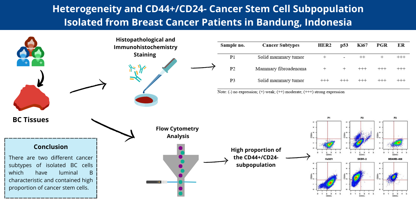
Breast cancer (BC) cells characteristics have played a crucial role in clinical strategies decision-making and patient outcome prediction. Although there have been several studies examining this, there are still relatively few that analyze stem cell subpopulations or protein biomarkers. The conducted research sought to elucidate breast cancer subtype characteristics based on immunohistochemical analysis and cancer stem cell presence based on flow cytometry analysis in three breast cancer samples from Bandung, Indonesia. A flow cytometry analysis was conducted to determine how much CD44/CD24 was expressed as a marker of cancer stem cell in isolated BC cells using various cell references, such as cell lines of SKBR-3, MDA-MB-468, and Hs587T. Immunohistochemistry analysis was carried out to investigate the expression of p53, HER2, PGR, Ki67, and ER in sample tissue sections. According to histological analysis, 2 samples were solid mammary carcinoma, and 1 sample was mammary fibroadenoma. Immunohistochemistry and histological analysis showed that all BC tissues are classified as Luminal B. In addition, a large percentage of CD44+/CD24- subpopulation was found in isolated BC cells (> 90 % in patients 1 and 3; > 60 % in patient 2). All samples showed similarity to Hs587T cell line characteristic. This study has successfully characterized solid mammary tumors and mammary fibroadenoma with luminal B characteristics. All specimens contained a high proportion of cancer stem cells.
HIGHLIGHTS
- The characteristics of breast cancer (BC) cells are essential factors in clinical strategy decision-making and patient outcome prediction
- Solid mammary tumor and mammary fibroadenoma were the cancer subtypes of isolated BC tissues from 3 different patients. The cancer subtypes found in 3 different patient’s isolated BC were solid mammary tumor and mammary fibroadenoma
- BC tissues from all 3 patients expressed different level of HER2, p53, Ki67, PGR and ER. Based on markers expressed, those BC tissues were classified as luminal B tumors
- Based on flow cytometry analysis, all BC tissues contained a high proportion of cancer stem cells indicated by the high proportion of CD44+/CD24- subpopulation
GRAPHICAL ABSTRACT
Downloads
References
J Ferlay, I Soerjomataram, R Dikshit, S Eser, C Mathers, M Rebelo, DM Parkin, D Forman and F Bray. Cancer incidence and mortality worldwide: Sources, methods and major patterns in GLOBOCAN 2012. Int. J. Canc. 2015; 136, E359-E386.
G Turashvili and E Brogi. Tumor heterogeneity in breast cancer. Front. Med. 2017; 4, 227.
MS Donepudi, K Kondapalli, SJ Amos and P Venkanteshan. Breast cancer statistics and markers. J. Canc. Res. Therapeut. 2014; 10, 506-11.
K Polyak. Heterogeneity in breast cancer. J. Clin. Investig. 2011; 121, 3786-8.
L Hutchinson. Challenges, controversies, breakthroughs. Nat. Rev. Clin. Oncol. 2010; 7, 669-70.
X Zhan, J Cheng, Z Huang, Z Han, B Helm, X Liu, J Zhang, TF Wang, D Ni and K Huang. Correlation analysis of histopathology and proteogenomics data for breast cancer. Mol. Cell. Proteomics 2019; 18, S37-S51.
B Weigelt, FC Geyer and JS Reis-Filho. Histological types of breast cancer: How special are they? Mol. Oncol. 2010; 4, 192-208.
C Mueller, A Haymond, JB Davis, A Williams and V Espina. Protein biomarkers for subtyping breast cancer and implications for future research. Expet. Rev. Proteomics 2018; 15, 131-52.
LNR Alves, DD Meira, LP Merigueti, MC Casotti, DP Ventorim, JFF Almeida, VPD Sousa, MC Sant’Ana, RGCD Cruz, LS Louro, GM Santana, TES Louro, RE Salazar, DRCD Silva, AS Siqueira Zetum, RSR Trabach, FIV Errera, F Paula, EVWD Santos, EFD Carvalho and ID Louro. Biomarkers in breast cancer: An old story with a new end. Genes 2023; 14, 1364.
AA Onitilo, JM Engel, RT Greenlee and BN Mukesh. Breast cancer subtypes based on ER/PR and Her2 expression: Comparison of clinicopathologic features and survival. Clin. Med. Res. 2009; 7, 4-13.
S Palomeras, S Ruiz-Martínez and T Puig. Targeting breast cancer stem cells to overcome treatment resistance. Molecules 2018; 23, 2193.
MJ Duffy, N Harbeck, M Nap, R Molina, A Nicolini, E Senkus and F Cardoso. Clinical use of biomarkers in breast cancer: Updated guidelines from the European Group on Tumor Markers (EGTM). Eur. J. Canc. 2017; 75, 284-98.
C Constantinou, S Papadopoulos, E Karyda, A Alexopoulos, N Agnanti, A Batistatou and H Harisis. Expression and clinical significance of claudin-7, PDL-1, PTEN, c-Kit, c-Met, c-Myc, ALK, CK5/6, CK17, p53, EGFR, Ki67, p63 in triple-negative breast cancer - a single centre prospective observational study. Vivo 2018; 32, 303-11.
CM Perou, T Sørlie, MB Eisen, MVD Rijn, SS Jeffrey, CA Rees, JR Pollack, DT Ross, H Johnsen, LA Akslen, Ø Fluge, A Pergamenschikov, C Williams, SX Zhu, PE Lønning, AL Børresen-Dale, PO Brown and D Botstein. Molecular portraits of human breast tumours. Nature 2000; 406, 747-52.
J Holm, L Eriksson, A Ploner, M Eriksson, M Rantalainen, J Li, P Hall and K Czene. Assessment of breast cancer risk factors reveals subtype heterogeneity. Canc. Res. 2017; 77, 3708-17.
SK Yeo and JL Guan. Breast cancer: Multiple subtypes within a tumor? Trends Canc. 2017; 3, 753-60.
W Sun and J Yang. Functional mechanisms for human tumor suppressors. J. Canc. 2010; 1, 136-40.
M Varna, G Bousquet, LF Plassa, P Bertheau and A Janin. TP53 status and response to treatment in breast cancers. J. Biomed. Biotechnol. 2011; 2011, 284584.
MH Jang, HJ Kang, KS Jang, SS Paik and WS Kim. Clinicopathological analysis of CD44 and CD24 expression in invasive breast cancer. Oncol. Lett. 2016; 12, 2728-33.
DR Siregar and A Christoper. Prevalence and characteristics of breast cancer in young women. Sumatera Med. J. 2020; 3, 1-9.
I Widodo, EK Dwianingsih, T Aryandono and S Soeripto. Clinicopathological characteristic and prognostic significance of Indonesian triple negative breast cancer. Indonesian Biomed. J. 2019; 11, 286-92.
W Widowati, Y Heriady, DR Laksmitawati, DK Jasaputra, TL Wargasetia, R Rizal, FS Perdana, A Amalia, A Arlisyah, Z Khoiriyah, A Faried and M Subangkit. Isolation, characterization and proliferation of cancer cells from breast cancer patients. Acta Inform. Med. 2018; 26, 240-4.
NA Pham, A Morrison, J Schwock, S Aviel-Ronen, V Iakovlev, MS Tsao, J Ho and DW Hedley. Quantitative image analysis of immunohistochemical stains using a CMYK color model. Diagn. Pathol. 2007; 2, 8.
D Ponti, A Costa, N Zaffaroni, G Pratesi, G Petrangolini, D Coradini, S Pilotti, MA Pierotti and MG Daidone. Isolation and in vitro propagation of tumorigenic breast cancer cells with stem/progenitor cell properties. Canc. Res. 2005; 65, 5506-11.
X Jiao, AA Rizvanov, M Cristofanilli, RR Miftakhova and RG Pestell. Breast cancer stem cell isolation. In: J Cao (Ed.). Humana Press, New York, 2016, p.121-35.
AP Shi, ZM Fan, KW Ma, YF Jiang, L Wang, KW Zhang, SB Fu, N Xu and ZR Zhang. Isolation and characterization of adult mammary stem cells from breast cancer adjacent tissues. Oncol. Lett. 2017; 14, 2894-902.
SR Lakhani, IO Ellis, SJ Schnitt, PH Tan and MJVD Vijver. WHO classification of tumours of the breast. IARC Press, Lyon, France, 2012.
C Sheridan, H Kishimoto, RK Fuchs, S Mehrotra, P Bhat-Nakshatri, CH Turner, R Goulet, S Badve and H Nakshatri. CD44+/CD24− breast cancer cells exhibit enhanced invasive properties: An early step necessary for metastasis. Breast Canc. Res. 2006; 8, R59.
Downloads
Published
How to Cite
Issue
Section
License
Copyright (c) 2023 Walailak University

This work is licensed under a Creative Commons Attribution-NonCommercial-NoDerivatives 4.0 International License.


