
Correlation Among Tumor Size, Histopathological Grade and the Expression of RAC1 Protein with Breast Cancer Metastasis to Ipsilateral Axillary Lymph Nodes
DOI:
https://doi.org/10.48048/tis.2023.6672Keywords:
Breast cancer, Axillary lymph nodes, Tumor size, Histopathologic grade, RAC1Abstract
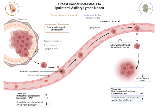
Breast cancer (BC) is the most common cancer affecting women worldwide. Mastectomy is the standard treatment for breast cancer. The purpose of axillary lymph nodes (ALN) dissection in mastectomy is to examine the status and the presence of metastasis in ALN. This study is an analytic observational cross-sectional study coupled with a categorical comparative study in 60 BC patient’s with- and without-ALN metastasis. The data that used in this study were histopathological data from patients medical record and immunohistochemistry (IHC) of RAC1 expression data. The immunohistochemistry was performed with a cutoff point based on the result of a receiver operating characteristics (ROC). This study combined BC tumor size, histopathological grade, and expression of RAC1 protein in correlation with known risk factors on metastasis to ALN. From 60 subjects, there were 63.3 % (38) ALN (+) and 36.7 % (22), ALN (−). BC of T2 tumor size, grade 2 and RAC1(−), the ALN metastasis chance was 0.6 %. T2 tumor size, grade 2 and RAC1(+), the ALN metastasis chance was 4.5 %. T2 tumor size, grade 3 and RAC1(−), the ALN metastasis chance was 3.0 %. T2 tumor, grade 3 and RAC1(+), the ALN metastasis chance was 18.8 %; T3 tumor size, grade 3 and RAC1(−), the ALN metastasis chance was 12.7 %. T3 tumor size, grade 3 and RAC1(+), the ALN metastasis chance was 52.2 %; T4b tumor size, grade 3, RAC1(−) and RAC1(+), the ALN metastasis chance was 40.8 and 83.8 %, respectively. We conclude that smaller tumor size and lower histopathological grading with RAC1(−) has lesser possibility to ALN metastasize.
HIGHLIGHTS
- Breast cancer (BC) is a malignant neoplasm originating from epithelial cells of the ducts and breast glands in the area of the lobules and terminal ducts known as the terminal duct lobular unit (TDLU) and the surgery performed is breast-conserving treatment or mastectomy that examine the status and the presence of metastasis in ALN
- RAC1 is a member of the RHO-GTPase family that plays an important role in the migration process, loss of adhesions between cells, and tumor metastasis also found in BC so it was used as parameter in this study
- Tumor size, histopathological grading, and the expression of RAC1 protein has correlation with with BC metastasis to the axillary lymph nodes that the smaller the tumor size, the lower the histopathological grading with RAC1(-)
GRAPHICAL ABSTRACT
Downloads
References
V Kumar, AK Abbas and C Jon. Aster: Robbins and cotran pathologic basis of disease. Elsevier, Amsterdam, Netherlands, 2020.
WHO, Breast Cancer, Available at: https://www.who.int/news-room/fact-sheets/detail/breast-cancer, accessed January 2023.
American Cancer Society. Breast cancer facts & figures 2013-2014. American Cancer Society, Inc, Atlanta, Georgia, 2013.
Perpustakaan Badan Kebijakan Pembangunan Kesehatan. Profil pengendalian penyakit dan penyehatan lingkungan tahun. Perpustakaan Badan Kebijakan Pembangunan Kesehatan, Jakarta, Indonesia, 2008.
International Agency for Research on Cancer, Available at: https://gco.iarc.fr/today/data/factsheets/ populations/360-indonesia-fact-sheets.pdf, accessed April 2021.
LH Sobin, MK Gospodarowicz and C Wittekind. TNM Classification of malignant tumours. John Wiley & Sons, Inc., New York, 2016.
VT DeVita, TS Lawrence and SA Rosenberg. DeVita, Hellman, and Rosenberg’s Cancer: Principles & practice of oncology. Lippincott Williams & Wilkins, Pennsylvania, 2008.
AT Mancino. Diseases of the breast. Ann. Surg. 2001; 233, 594.
C Eiden and H Ungefroren. The ratio of RAC1B to RAC1 expression in breast cancer cell lines as a determinant of epithelial/mesenchymal differentiation and migratory potential. Cells 2021; 10, 351.
Y Heriady, D Achmad, BS Hernowo, A Faried, D Ismono and D Hilmanto. Expression of the RAC1, RHOA and CXCR4 proteins and their interaction as risk factors for infiltration to the nipple areola complex in operable breast carcinoma. Breast Canc. 2019; 26, 172-9.
S Sandoughdaran, M Malekzadeh and MEM Eismael. Frequency and predictors of axillary lymph node metastases in iranian women with early breast cancer. Asian Pac. J. Canc. Prev. 2018; 19, 1617-20.
AJ Ridley. Rho GTPases and cell migration. J. Cell Sci. 2001; 114, 2713-22.
TC Tu, M Nagano, T Yamashita, H Hamada, K Ohneda, K Kimura and O Ohneda. A chemokine receptor, CXCR4, which is regulated by hypoxia-inducible factor 2α, is crucial for functional endothelial progenitor cells migration to ischemic tissue and wound repair. Stem Cell. Dev. 2016; 25, 266-76.
DR Welch and DR Hurst. Defining the hallmarks of metastasis. Canc. Res. 2019; 79, 3011-27.
P Chand, S Singh, G Singh, S Kundal and A Ravish. A study correlating the tumor site and size with the level of axillary lymph node involvement in breast cancer. Niger. J. Surg. 2020; 26, 9-15.
NI Hadi and Q Jamal. Comparison of clinicopathological characteristics of lymph node positive and lymph node negative breast cancer. Pakistan J. Med. Sci. 2016; 32, 863-8.
S Yenidunya, R Bayrak and H Haltas. Predictive value of pathological and immunohistochemical parameters for axillary lymph node metastasis in breast carcinoma. Diagn. Pathol. 2011; 6, 18.
AA Mohammed. Predictive factors affecting axillary lymph node involvement in patients with breast cancer in Duhok: Cross-sectional study. Ann. Med. Surg. 2019; 44, 87-90.
Downloads
Published
How to Cite
Issue
Section
License
Copyright (c) 2023 Walailak University

This work is licensed under a Creative Commons Attribution-NonCommercial-NoDerivatives 4.0 International License.


