
Characterization of Microencapsulation Human Adipose Tissue Mesenchymal Stem Cells (hAT-MSCs) and Nanoparticle of Conditioned Medium of AT-MSCs
DOI:
https://doi.org/10.48048/tis.2023.6425Keywords:
Conditioned medium, Human adipose tissue mesenchymal stem cell, Microencapsulation, NanoparticlesAbstract
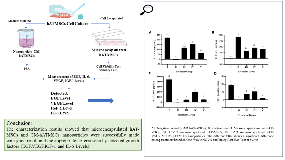
This study aims to characterize the microencapsulated type of human Adipose Tissue Mesenchymal Stem Cells (hAT-MSCs) and nanoparticles containing a conditioned medium of hAT-MSCs (CM-hATMSCs). The hAT-MSCs microencapsulation was measured the cells viability on 1st, 7th, and 14th days, by 3-(4,5-dimethylthiazol-2-yl)-5-(3-carboxymethylphenyl)-2-(4-sulfophenyl)-2H-tetrazolium (MTS) assay, stability test was performed using magnetic stirrer at a speed of 300, 700, 1100, and 1200 rpm (revolutions per minutes) and applied on hAT-MSCs microencapsulation. The particle size of CM-hATMSCs nanoparticles were measured by particle size analyzer (PSA) and the growth factor levels namely Epidermal Growth Factor (EGF), Interleukin-6 (IL-6), Vascular Endothelial Growth Factor (VEGF), Insulin-Like Growth Factor-1 (IGF-1) were investigated using ELISA method. In the 14th day showed decreasing of cell viability percentage from 90 - 100% to 50% in both of 1.5×10⁵ and 3×10⁵ cells hAT-MSCs microencapsulation. CM-hATMSCs microencapsulation are quite stable and has a fairly good mechanical resistance when tested for stability using rotation at a speed of 1,200 rpm only damages 10 % of it. The CM-hATMSCs nanoparticles collected from hAT-MSCs growth medium in PSA test have an average particle size of 141 nm. Based on measurement of growth factor level, hAT-MSCs microencapsulation (3×10⁵ cells) and CM-hATMSCs nanoparticles showed secretion of growth factors of EGF, IL-6, VEGF, and IGF-1 compared to positive controls. In summary, hAT-MSCs microencapsulation formulation using alginate showed good product based on the viability, stability, structure, and growth factor secretion ability. The characterization results showed that microencapsulated hAT-MSCs and CM-hATMSCs nanoparticles were successfully synthesized with the appropriate criteria.
HIGHLIGHTS
- Stem cell therapy using Human ATMSCs (hAT-MSCs) has the potential to accelerate wound healing on the skin both directly and indirectly
- hMSC transplantation is affected by environment and inflammation which can trigger unwanted cell differentiation, so the solution is to use the nanoparticle method (CM-hATMSC) as a drug mobilization system
- hAT-MSCs and CM-hATMSCs display high secretion of growth factors namely EGF, IL-6, VEGF, and IGF-1
- hAT-MSCs and CM-hATMSCs characterization was successfully synthesized according to the criteria
GRAPHICAL ABSTRACT
Downloads
References
A Meiliana, NM Dewi and A Wijaya. Stem cell therapy in wound healing and tissue regeneration. Indones. Biomed. J. 2016; 8, 61-70.
A Meiliana, NM Dewi and A Wijaya. Mesenchymal stem cells manage endogenous tissue regeneration. Indones. Biomed. J. 2016; 8, 71-90.
X Guo, C Schaudinn, U Blume-Peytavi, A Vogt and F Rancan. Effects of adipose-derived stem cells and their conditioned medium in a human ex vivo wound model. Cells 2022; 11, 1-24.
Y Lina and A Wijaya. Adipose-derived stem cells for future regenerative system medicine. Indones. Biomed. J. 2012; 4, 59-72.
A Krawczenko and A Klimczak. Adipose tissue-derived mesenchymal stem/stromal cells and their contribution to angiogenic processes in tissue regeneration. Int. J. Mol. Sci. 2022; 23, 1-20.
R Noverina, W Widowati, W Ayuningtyas, D Kurniawan, E Afifah, DR Laksmitawati, R Rinendyaputri, R Rilianawati, A Faried, I Bachtiar and EF Wirakusumah. Growth factors profile in conditioned medium human adipose tissue-derived mesenchymal stem cells (CM-hATMSCs). Clin. Nutr. Exp. 2019; 24, 34-44.
BS Park, KA Jang, JH Sung, JS Park, YH Kwon, KJ Kim and WS Kim. Adipose-derived stem cells and their secretory factors as a promising therapy for skin aging. Dermatol. Surg. 2008; 34, 1323-6.
L Bussche and GRVD Walle. Peripheral blood-derived mesenchymal stromal cells promote angiogenesis via paracrine stimulation of vascular endothelial growth factor secretion in the equine model. Stem Cells Transl. Med. 2014; 3, 1514-25.
L Bussche, RM Harman, BA Syracuse, EL Plante, YC Lu, TM Curtis, M Ma and GRV Walle. Microencapsulated equine mesenchymal stromal cells promote cutaneous wound healing in vitro. Stem Cell Res. Ther. 2015; 6, 1-15.
A Roffi, N Nakamura, M Sanchez, M Cucchiarini and G Filardo. Injectable systems for intra-articular delivery of mesenchymal stromal cells for cartilage treatment: A systematic review of preclinical and clinical evidence. Int. J. Mol. Sci. 2018; 19, 1-19.
S Lee, E Choi, MJ Cha and KC Hwang. Cell adhesion and long-term survival of transplanted mesenchymal stem cells: A prerequisite for cell therapy. Oxid. Med. Cell Longev. 2015; 2015, 1-9.
A Waheed, MAJ Mazumder, A Al-Ahmed, P Roy and N Ullah. Cell encapsulation. Funct. Biopolym. 2019; 2019, 377-27.
JA Barminko. 2012, Encapsulated mesenchymal stromal cells for spinal cord injury repair. Ph. D. Dissertation. Ruoole tgers University, New Brunswick, Canada.
B Kupikowska-Stobba and D Lewińska. Polymer microcapsules and microbeads as cell carriers for: In vivo biomedical applications. Biomater. Sci. 2020; 8, 1536-74.
R Martien, Adhyatmika, IDK Irianto, V Farida and DP Sari. Technology developments nanoparticles as drug delivery systems. Majalah Farmaseutik. 2012; 8, 133-44.
W Widowati, R Noverina, W Ayuningtyas, D Kurniawan, S Arumwardana, HSW Kusuma and A Faried. Potential of conditioned medium of hATMSCs in aging cells model. Hayati J. Biosci. 2022; 29, 378-88.
W Widowati, E Afifah, T Mozef, F Sandra, R Rizal, A Amalia, Y Arinta, I Bachtiar and H Murti. Effects of insulin-like growth factor-induced Wharton jelly mesenchymal stem cells toward chondrogenesis in an osteoarthritis model. Iran J. Basic Med. Sci. 2018; 21, 745-52.
D Rahmat, W Widowati, A Faried, IM Nainggolan, D Priyandoko, A Budiati, TM Zakaria and E Afifah. Microencapsulation of Indonesian polymer biodiversity in Wharton’s jelly mesenchymal stem cell (WJMSC). Int. J. Appl. Pharm. 2022; 14, 91-4.
W Widowati, DK Jasaputra, SB Sumitro, MA Widodo, T Mozef, R Rizal, HSW Kusuma, DR Laksmitawati, H Murti, I Bachtiar and A Faried. Effect of interleukins (Il-2, IL-15, IL-18) on receptors activation and cytotoxic activity of natural killer cells in breast cancer cell. African Health Sci. 2020; 20, 822-32.
W Widowati, H Murti, DK Jasaputra, SB Sumitro, MA Widodo, N Fauziah, M Maesaroh and I Bachtiar. Selective cytotoxic potential of IFN-γ and TNF-α on breast cancer cell lines (T47D and MCF7). Asian J. Cell Biol. 2016; 11, 1-12.
W Widowati, L Wijaya, H Murti, H Widyastuti, D Agustina, DR Laksmitawati, N Fauziah, SB Sumitro, MA Widodo and I Bachtiar. Conditioned medium from normoxia (WJMSCs-norCM) and hypoxia-treated WJMSCs (WJMSCs-hypoCM) in inhibiting cancer cell proliferation. Biomark. Genom. Med. 2015; 7, 8-17.
R Negrulj, A Mooranian, N Chen-Tan, HS Al-Sallami, M Mikov, S Golocorbin-Kon and H Al-Salami. Swelling, mechanical strength, and release properties of probucol microcapsules with and without a bile acid, and their potential oral delivery in diabetes. Artif. Cells Nanomed. Biotechnol. 2016; 44, 1290-7.
R Mahou, RPH Meier, LH Bühler and C Wandrey. Alginate-poly(ethylene glycol) hybrid microspheres for primary cell microencapsulation. Mater. 2014; 7, 275-86.
D Rahmat, W Widowati, A Faried, IM Nainggolan, D Priyandoko, A Budiati, TM Zakaria and E Afifah. Characterization of nanoparticles conditioned medium adipose tissue mesenchymal stem cell (CM-ATMSC). Int. J. Appl. Pharm. 2022; 14, 104-6.
S Farid, S Ameen, S Sharif, M Tariq, IA Kundi, O Sahin, MS Sayyad and IU Khan. Facile solvothermal syntheses of isostructural lanthanide (III) formates: Photocatalytic, photoluminescent chemosensing properties, and proficient precursors for metal oxide nanoparticles. J. Coord. Chem. 2021; 74, 1700-19.
W Widowati, L Darsono, J Suherman, N Fauziah, M Maesaroh and P Erawijantari. Anti-inflammatory effect of mangosteen (Garcinia mangostana l.) peel extract and its compounds in LPS-induced RAW264.7 cells. Nat. Prod. Sci. 2016; 22, 147-53.
W Widowati, H Widyastuti, H Murti, DR Laksmitawati, M Maesaroh, SB Sumitro, A Widodo and I Bachtiar. Interleukins and VEGF secretome of human Wharton’s jelly mesenchymal stem cells-conditioned medium (hWJMSCs-CM) in different passages and oxygen tensions. Biosci. Res. 2017; 14, 776-87.
Y Lina, GS Lawrence and A Wijaya. Association of free fatty acid (FFA), fatty acid binding protein (FABP) and adiponectin with tumor necrosis factor-alpha (TNF-alpha) and interleukin-6 (IL-6) among obese non diabetic males. Indones. Biomed. J. 2009; 1, 31-7.
D Sadoughi, MA Edalatmanesh and R Rahbarian. Protective effect of curcumin on quality parameters ofsperm and testicular tissue alterations in alloxan-induced diabetic rats as animal model. Indones. Biomed. J. 2019; 11, 240-6.
PD Vos, HA Lazarjani, D Poncelet and MM Faas. Polymers in cell encapsulation from an envelopedcell perspective. Adv. Drug Deliv. Rev. 2014; 67, 15-34.
GAP Juárez, M Spasojevic, MM Faas and PD Vos. Immunological and technical considerationsin application of alginate-based microencapsulation systems. Front. Bioeng. Biotechnol. 2014; 2, 1-15.
G Lv, Z Sun, S Li, W Yu, Y Xi, Y Zhang, H Xiw, X Li, W Wang and X M. Permeability changes ofthe cell-contained microcapsules visualized by confocal laser scanning microscope. J. Biomed. Mater. Res. A 2009; 90, 773-83.
J Xi and M Talaat. Nanoparticle deposition in rhythmically moving acinar models with interalveolar septal apertures. Nanomaterials 2019; 9, 1-18.
Y Aghazadeh and MC Nostro. Cell therapy for type 1 diabetes: Current and future strategies. Curr. Diab. Rep. 2017; 17, 1-9.
R Krishnan, M Alexander, L Robles, CE Foster and JR Lakey. Islet and stem cell encapsulation for clinical transplantation. Rev. Diab. Stud. 2014; 11, 84-1.
Downloads
Published
How to Cite
Issue
Section
License
Copyright (c) 2023 Walailak University

This work is licensed under a Creative Commons Attribution-NonCommercial-NoDerivatives 4.0 International License.


