
Cellular Development and Ultrastructural Characteristics of Halymenia durvillei Bory de Saint-Vincent, 1828
DOI:
https://doi.org/10.48048/tis.2027.14471Keywords:
Halymenia durvillei, Red algae, Cellular, Ultrastructure, Chloroplast, Phycobilisome, Development, Halymenia durvillei, Red algae, Cellular, Ultrastructure, Chloroplast, Phycobilisome, DevelopmentAbstract
Halymenia durvillei Bory de Saint-Vincent, 1828 is a marine red alga with potential relevance for aquaculture and biomass production. Samples were collected from a Coastal Aquaculture Research Center in Thailand, and cultivated in culture tanks with natural seawater for up to 3 months. This study examined the morphological and ultrastructural changes in H. durvillei thalli across different cultivation periods to assess developmental progression and determine an optimal harvest time for the most mature growth stage to maximize biomass production. Molecular identification based on the rbcL gene sequencing confirmed the taxonomic identity of the studied alga, showing 99% sequence similarity to H. durvillei. Light microscopy (LM) and transmission electron microscopy (TEM) revealed progressive increases in thallus thickness and pronounced changes in cellular organization with increasing cultivation period. Cell size increased markedly, and the differences in outer cortical cells became significantly apparent with cultivation time. Each cell contained a single large chloroplast with well-developed thylakoids, phycobilisomes, pyrenoids, and plastoglobules. In addition, floridean starch granules and lipid droplets were observed in the cytoplasm and increased with the cultivation period. These ultrastructural changes indicate enhanced development of chloroplast-associated storage systems, reflecting increased accumulation of carbohydrate and lipid reserves. After 3 months of cultivation, thalli exhibited the largest cell size and the most pronounced development of chloroplast-associated storage structures, suggesting advanced plastid maturation and metabolic readiness. Overall, a cultivation period of approximately 3 months was optimal for obtaining mature thalli with enhanced storage capacity. H. durvillei is a high-value red seaweed with broad potential applications in the food, pharmaceutical, and biotechnological industries.
HIGHLIGHTS
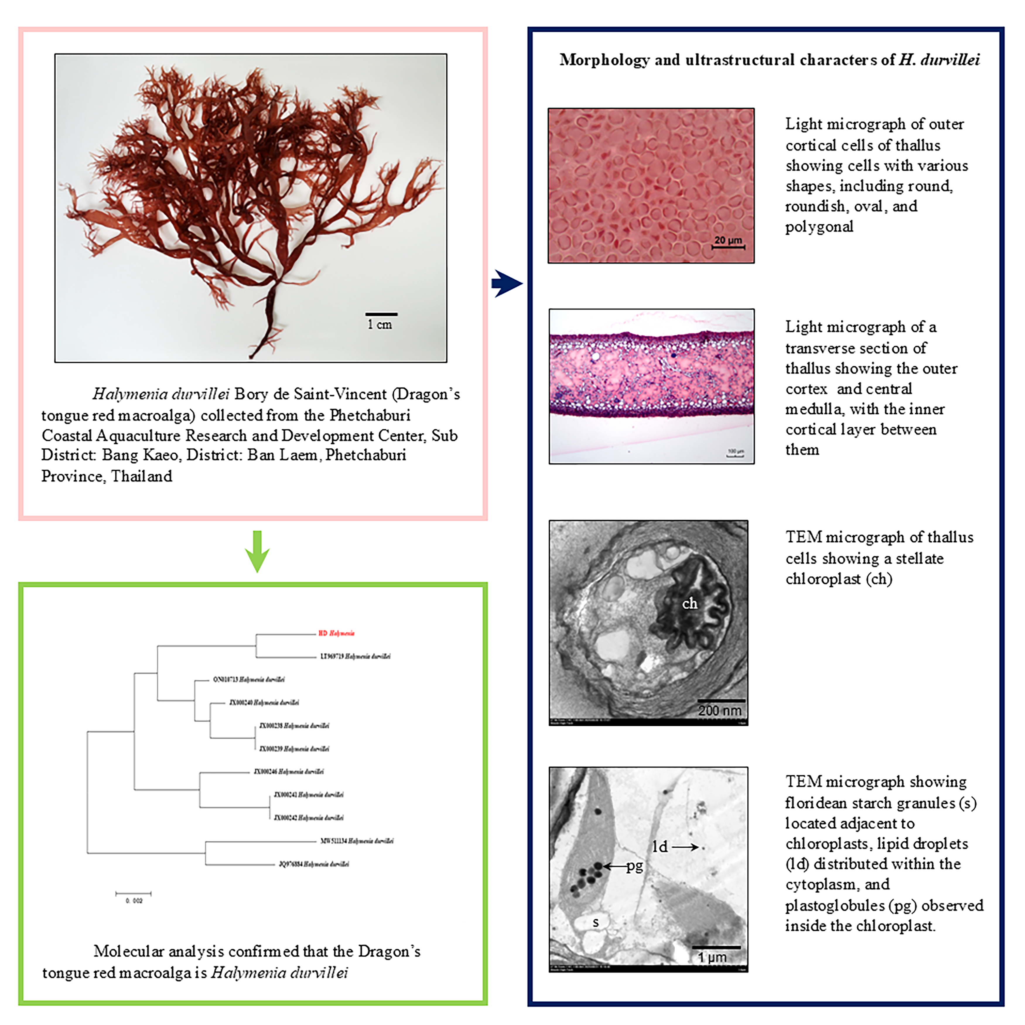
- The molecular analysis of Dragon’s tongue red macroalgae confirmed the identity of Halymenia durvillei based on rbcL gene sequencing. The morphological and ultrastructural characters of durvillei were studied using light and transmission electron microscopy.
- The morphological and ultrastructural characters of durvillei were studied using light and transmission electron microscopy.
- The outer cortical cell length increased from 9.57 mm to 12.05 mm, width from 7.38 to 9.84 mm, and thallus thickness from 340.73 to 558.10 mm of durvillei during the 0 - 3 months cultivation period.
- Each cell contained a large stellate chloroplast with plastoglobules, while floridean starch granules adjacent to the chloroplast increased with the cultivation period.
GRAPHICAL ABSTRACT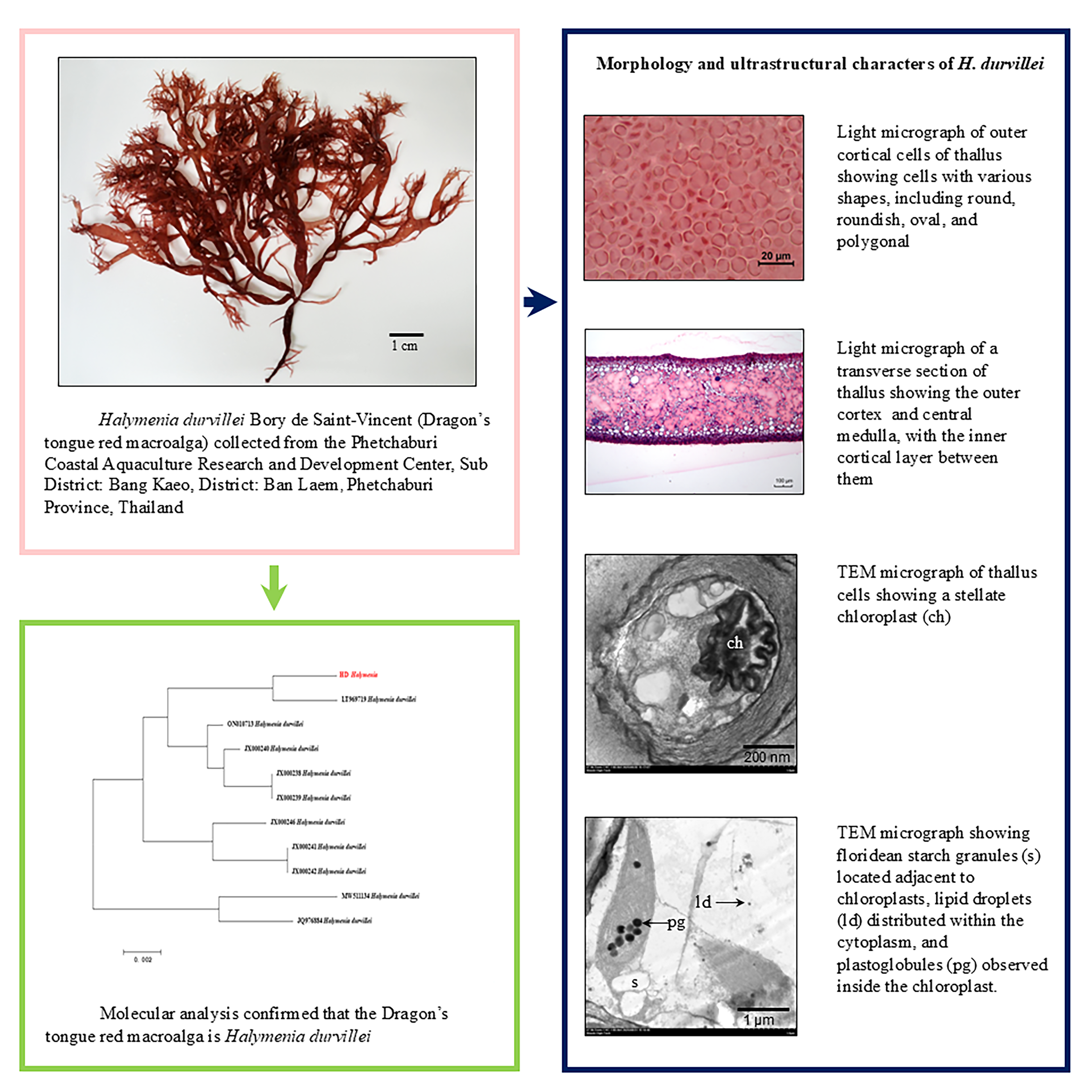
Downloads
References
JS Zaneveld. The utilization of marine algae in tropical South and East Asia. Economic Botany 1959; 13(2), 89-131.
EE Mon. The morphology and distribution of Halymenia durvillei Bory de Saint-Vincent (Halymeniales, Rhodophyta) from Myanmar. Journal of Aquatic Science and Marine Biology 2018; 1(4), 15-25.
ADR N’Yeurt. Marine algae from the Suva lagoon and reef, Fiji. Australian Systematic Botany 2001; 14(5), 689-869.
G de Smedt, O de Clerck, F Leliaert, E Coppejans and LM Liao. Morphology and systematics of the genus Halymenia C. Agardh (Halymeniales, Rhodophyta) in the Philippines. Nova Hedwigia 2001; 73(3-4), 293-322.
S Kawaguchi, S Shimada, T Abe and R Terada. Morphological and molecular phylogenetic studies of a red alga, Halymenia durvillei, (Halymeniaceae, Halymeniales) from Indo-Pacific. Coastal Marine Sciences 2006; 30(1), 201-208.
RE Lee. Phycology. Cambridge University Press, New York, 2008, p. 16-93.
DE Schindel and SE Miller. DNA barcoding a useful tool for taxonomists. Nature 2005; 435(7038), 17.
BE Clarkston and GW Saunders. A comparison of two DNA barcode markers for species discrimination in the red algal family Kallymeniaceae (Gigartinales, Florideophyceae), with a description of Euthora timburtonii sp. nov. Botany 2010; 88(2), 119-131.
DW Freshwater and J Rueness. Phylogenetic relationships of some European Gelidium (Gelidiales, Rhodophyta) species, based on rbcL nucleotide sequence analysis. Phycologia 1994; 33(3), 187-194.
B Gavio and S Fredericq. Grateloupia turuturu (Halymeniaceae, Rhodophyta) is the correct name of the non-native species in the Atlantic known as Grateloupia doryphora. European Journal of Phycology 2002; 37(3), 349-359.
MS Kim, SY Kim and W Nelson. Symphyocladia lithophila sp. nov. (Rhodomelaceae, Ceramiales), a new Korean red algal species based on morphology and rbcL sequences. Botanica Marina 2010; 53(3), 233-241.
AR Sherwood and GG Presting. Universal primers amplify a 23S rDNA plastid marker in eukaryotic algae and cyanobacteria. Journal of Phycology 2007; 43(3), 605-608.
HW Wang, S Kawaguchi, T Horiguchi and M Masuda. Reinstatement of Grateloupia catenata (Rhodophyta, Halymeniaceae) on the basis of morphology and rbcL sequences. Phycologia 2000; 39(3), 228-237.
JL Godínez-Ortega, P Snoeijs, D Robledo, Y Freile-Pelegrín and M Pedersén. Growth and pigment composition in the red alga Halymenia floresii cultured under different light qualities. Journal of Applied Phycology 2008; 20, 253-260.
JB Barufi, FL Figueroa and EM Plastino. Effects of light quality on reproduction, growth and pigment content of Gracilaria birdiae (Rhodophyta: Gracilariales). Scientia Marina 2015; 79(1), 15-24.
NAM Rula, ET Ganzon-Fortes, MJR Pante and GC Trono Jr. Influence of light, water motion, and stocking density on the growth and pigment content of Halymenia durvillei (Rhodophyceae) under laboratory conditions. Journal of Applied Phycology 2021; 33, 2367-2377.
A Balkrishna, U Agarwal, S Shrivastava, S Chaudhary and V Arya. Therapeutic potential and applications of red algae: A comprehensive scientific review. Phytochemistry Reviews 2026; 25, 821-852.
II Osovskaya and EA Krivorotova. Properties of agarose - a polysaccharide from marine red algae. Fibre Chemistry 2024; 55(6), 369-372.
A Chaiwichien, T Samrit, S Osotprasit, P Kueakhai, P Sobhon, K Meemon, N Niamnont, P Manohong, K Pranweerapaiboon, M Tamtin and N Changklungmoa. Evaluation of toxicity and anti-oxidation activity of the extracts from Halymenia durvillei. Trends in Sciences 2022; 19(6), 3032.
P Vivithanaporn, T Siangcham, V Tanawoot, R Settacomkul, K Pranweerapaiboon, K Meemon, N Niamnont, M Tamtin, P Sobhon and K Sangpairoj. Apoptotic and autophagic cell death effects of the hexane extract of tropical marine algae Halymenia durvillei against human glioblastoma cells: In vitro and in silico studies. Trends in Sciences 2024; 21(2), 7157.
JJ Doyle and JL Doyle. A rapid DNA isolation procedure for small quantities of fresh leaf tissue. Phytochemical Bulletin 1987; 19, 11-15.
M Johnson, I Zaretskaya, Y Raytselis, Y Merezhuk, S McGinnis and TL Madden. NCBI BLAST: A better web interface. Nucleic Acids Research 2008; 36(2), W5-W9.
JR Harris. Electron microscopy in biology: A practical approach. Oxford University Press, New York, 1991, p. 1-37.
MA Hayat. Principles and techniques of electron microscopy: Biology applications. Cambridge University Press, New York, 2000, p. 85-138.
BE Juniper, GC Cox, AJ Gilchrist and PR Williams. Techniques for plant electron microscopy. Holywell Press, Oxford, England, 1970, p. 1-19.
DC Pease. Histological techniques for electron microscopy. Academic Press, New York, 1964, p. 260-261.
CE McClung. Handbook of microscopical technique: For workers in animal and plant tissues. Hoeber, New York, 1950, p. 84-85.
RJV Pulvertaft. McClung’s handbook of microscopical technique. Journal of Clinical Pathology 1951; 4(4), 497.
JJ Bozzola and LD Russell. Electron microscopy: Principles and techniques for biologists. Jones & Bartlett Publishers, Boston, 1992, p. 112-114.
M Hoppert and A Holzenburg. Electron microscope in microbiology. Bios Scientific Publishers, Oxford, England, 1998, p. 55-57.
Y Paopun, P Thanomchat, C Roopkham, P Umroong, W Pan-Utai, P Satmalee, P Kosawatpat, B Thongdang and M Tamtin. Structural development of marine green alga (Ulva rigida C. Agardh, 1823) during cultivation. Trends in Sciences 2023; 20(8), 6747.
CAA de Azevedo, V Cassano and MC Oliveira. Diversity of branched Halymenia (Halymeniales, Rhodophyta) species on the Brazilian coast: Molecular and morphological analyses reveal three new species. Phycologia 2016; 55(4), 431-444.
JJ Hernandez-Kantun, AR Sherwood, R Riosmena-Rodriguez, JM Huisman and O de Clerck. Branched Halymenia species (Halymeniaceae, Rhodophyta) in the Indo-Pacific region, including descriptions of Halymenia hawaiiana sp. nov. and H. tondoana sp. nov. European Journal of Phycology 2012; 47(4), 421-432.
YW Sheng, W Zhang, D Zhao and HW Wang. A morphological and molecular assessment of the genus Sinotubimorpha (Halymeniaceae, Rhodophyta). Journal of Systematics and Evolution 2012; 50(2), 146-152.
PS Dixon. Biology of the rhodophyta. Oliver & Boyd, Edinburgh, Scotland, 1973, p. 93-110.
LE Graham, JM Graham and LW Wilcox. Algae. Benjamin Cummings, San Francisco, 2009, p. 309-324.
S Augyte, NA Sims, K Martin, SV Wychen, B Panczak, H Alt, R Nelson and LML Laurens. Tropical red macroalgae cultivation with a focus on compositional analysis. Plants 2023; 12(20), 3524.
J Cotas, A Leandro, P Monteiro, D Pacheco, A Figueirinha, AMM Gonçalves, GJ da Silva and L Pereira. Seaweed phenolics: From extraction to applications. Marine drugs 2020; 18(8), 384.
V Stiger-Pouvreau, N Bourgougnon and E Deslandes. Carbohydrates from seaweeds. In: J Fleurence and I Levine (Eds.). Seaweed in health and disease prevention. Academic Press, New York, 2016, p. 223-274.
ZA Popper, G Michel, C Hervé, DS Domozych, WGT Willats, MG Tuohy, B Kloareg and DB Stengel. Evolution and diversity of plant cell walls: From algae to flowering plants. Annual Review of Plant Biology 2011; 62, 567-590.
N Ghassemi, A Poulhazan, F Deligey, F Mentink-Vigier, I Marcotte and T Wang. Solid-state NMR investigations of extracellular matrixes and cell walls of algae, bacteria, fungi, and plants. Chemical Reviews 2022; 122, 10036-10086.
I Mine, K Yuasa, M Uesugi, S Sekida and K Okuda. Band growth and localization of vesicle exocytosis in the red alga Antithamnion nipponicum (Ceramiales). European Journal of Phycology 2011; 46(3), 263-270.
J Yang, S Chang, L Li, X Ma, H Han, Z Liu and J Zhu. Structural insights into cell wall-related vesicle secretion with different mechanisms. Plants 2026; 15(4), 660.
KA Pyke. Plastid division and development. The Plant Cell 1999; 11(4), 549-556.
ZL Bouzon, C Simioni and EC Schmidt. The structure and morphology of red algae chloroplasts. In: MF Hohmann-Marriott (Ed.). The Structural Basis of Biological Energy Generation. Springer, Dordrecht, Netherlands, 2014, p. 295-308.
K Solymosi. Plastid structure, diversification and interconversions I. Algae. Current Chemical Biology 2012; 6(3), 167-186.
L Barsanti and P Gualtieri. Algae: Anatomy, biochemistry, and biotechnology. CRC Press, Boca Raton, 2014, p. 118-122.
AR Grossman, MR Schaefer, GG Chiang and JL Collier. The phycobilisome, a light-harvesting complex responsive to environmental conditions. Microbiological Reviews 1993; 57(3), 725-749.
S Sekar and M Chandramohan. Phycobiliproteins as a commodity: Trends in applied research, patents and commercialization. Journal of Applied Phycology 2008; 20, 113-136.
H Nacir and C Bréhélin. When proteomics reveals unsuspected roles: The plastoglobule example. Frontiers in Plant Science 2013; 4, 114.
PK Lundquist. Chromoplast differentiation: A central role for plastoglobule lipid droplets comes into focus. New Phytologist 2003; 237, 1483-1485.
JN Lohscheider and CR Bártulos. Plastoglobules in algae: A comprehensive comparative study of the presence of major structural and functional components in complex plastids. Marine Genomics 2016; 28, 127-136.
A Converti, AA Casazza, EY Ortiz, P Perego and MD Borghi. Effect of temperature and nitrogen concentration on the growth and lipid content of Nannochloropsis oculata and Chlorella vulgaris for biodiesel production. Chemical Engineering and Processing: Process Intensification 2009; 48(6), 1146-1151.
BM Long, Y Matsuda and JV Moroney. Algal chloroplast pyrenoids: Evidence for convergent evolution. Proceedings of the National Academy of Sciences 2024; 121(14), e2402546121.
A Wahid and MA Khan. Viruses bacteria and thalloid organisms. Higher Education Commission, Islamabad, Pakistan, 2006, p. 72-77.
HC Bold and MJ Wynne. Introduction to the algae: Structure and reproduction. Prentice-Hall Inc, New Jersey, 1978, p. 504-505.
R Viola, P Nyvall and M Pedersén. The unique features of starch metabolism in red algae. Proceedings of the Royal Society B: Biological Sciences 2001; 268(1474), 1417-1422.
Q Hu, M Sommerfeld, E Jarvis, M Ghirardi, M Posewitz, M Seibert and A Darzins. Microalgal triacylglycerols as feedstocks for biofuel production: Perspectives and advances. The Plant Journal 2008; 54(4), 621-639.
Q Al-Abdallah, BT Nixon and JR Fortwendel. The enzymatic conversion of major algal and cyanobacterial carbohydrates to bioethanol. Frontiers in Energy Research 2016; 4, 36.
T Li, M Gargouri, J Feng, JJ Park, D Gao, C Miao, T Dong, DR Gang and S Chen. Regulation of starch and lipid accumulation in a microalga Chlorella sorokiniana. Bioresource Technology 2015; 180, 250-257.
Downloads
Published
How to Cite
Issue
Section
License
Copyright (c) 2026 Walailak University

This work is licensed under a Creative Commons Attribution-NonCommercial-NoDerivatives 4.0 International License.