
Understanding the Blood-Brain Barrier (BBB) with MRI Techniques and Its Implications in Neurodegenerative Diseases: An Overview
DOI:
https://doi.org/10.48048/tis.2024.8010Keywords:
Blood-brain barrier, Alzheimer’s disease, Parkinson’s disease, Multiple sclerosis, Dynamic contrast enanchement, Dynamic susceptibility contrast, Magnetic resonance imaging, Focused ultrasoundAbstract
The blood-brain barrier (BBB) stands as a critical guardian separating the central nervous system (CNS) from the systemic circulation. This comprehensive review explores the anatomical and functional components of the BBB and its association with the neurovascular unit (NVU), emphasizing its role in synaptic signaling and shielding the CNS from neurotoxic elements. Detailed discussions encompass MRI techniques like dynamic contrast enhancement (DCE) and arterial spin labeling (ASL) MRI, illuminating their significance in assessing BBB integrity and permeability. Various models and pharmacokinetic parameters utilized in imaging analysis offer insights into barrier permeability, aiding in the evaluation of neurodegenerative illnesses such as Alzheimer’s, Parkinson’s, and multiple sclerosis. Additionally, the study investigates the distinct characteristics of imaging protocols and their impact on BBB evaluation. Highlighting physiological conditions, the analysis discerns regional disparities in BBB permeability, shedding light on diverse microvascular architectures in healthy subjects. Conversely, in pathological states like Alzheimer’s, Parkinson’s, and multiple sclerosis, BBB dysfunction leads to a cascade of events facilitating the entry of harmful substances, exacerbating neurodegeneration. Imaging studies have unveiled distinct alterations in BBB permeability and perfusion, providing crucial insights into disease progression, notably preceding structural changes in Alzheimer’s and indicating localized disruptions in multiple sclerosis. This comprehensive exploration underscores the pivotal role of the BBB in maintaining CNS health and its intricate involvement in the pathogenesis of neurodegenerative disorders. While imaging techniques serve as promising tools for BBB assessment, further research is warranted to refine their diagnostic precision and differentiation abilities across neurological conditions.
HIGHLIGHTS
The central role of the blood-brain barrier (BBB) with its anatomical-functional components and the association with the neurovascular unit (NVU) represent the heart of this study whose aim is to evaluate and be able to grasp the pathological alterations of permeability and perfusion that suggest the existence of neurodegenerative diseases such as Alzheimer’s, Parkinson's and multiple sclerosis. Different MRI techniques that are used to evaluate BBB alterations such as dynamic contrast enhancement (DCE), MRI with arterial spin labeling (ASL) and various pharmacokinetic models and parameters will be discussed through a literature review in order to provide the radiologist and clinician clear indications regarding the main techniques suggested in the evaluation of this important anatomical-functional unit.
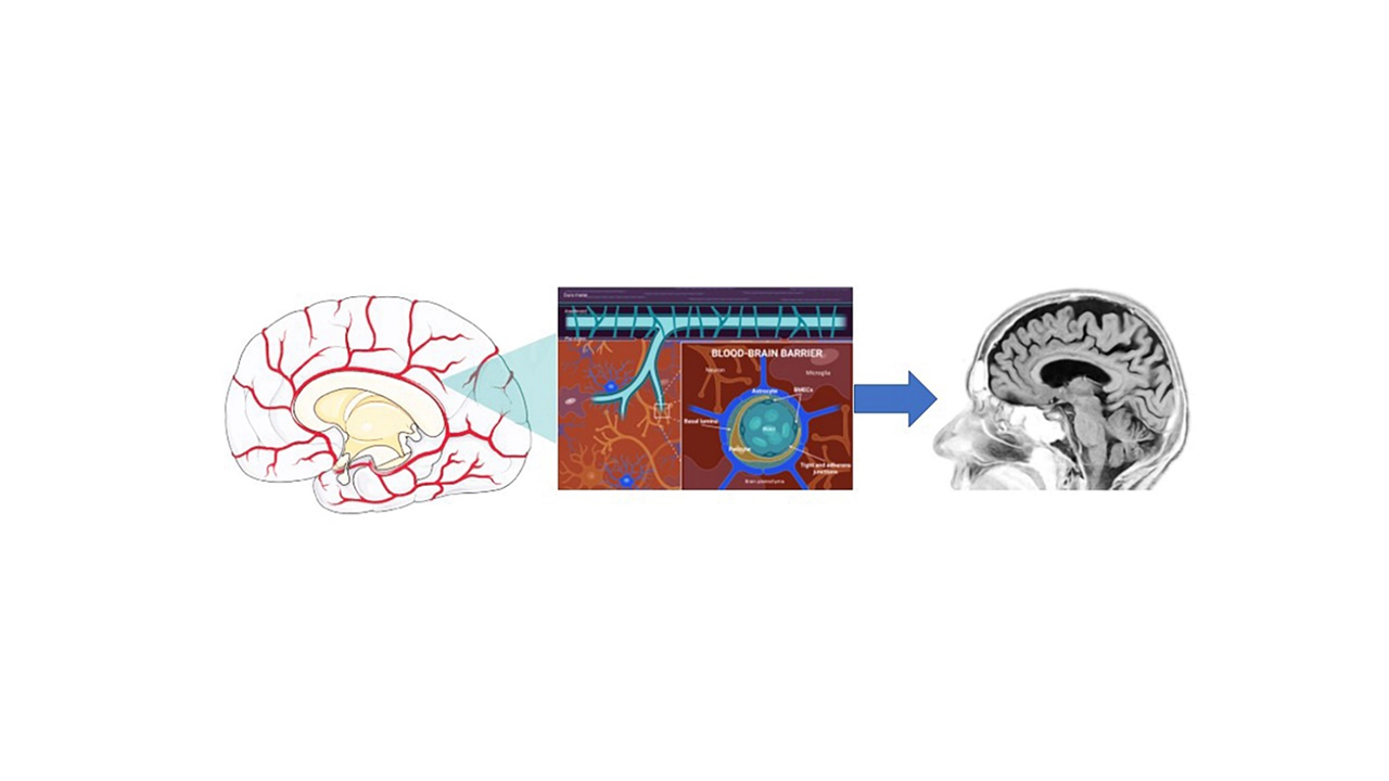
GRAPHICAL ABSTRACT
Downloads
References
I Wilhelm, A Nyul-Toth, M Suciu, A Hermenean and IA Krizbai. Heterogeneity of the blood-brain barrier. Tissue Barriers 2016; 4, e1143544.
PM Carvey, B Hendey and AJ Monahan. The blood-brain barrier in neurodegenerative disease: A rhetorical perspective. J. Neurochem. 2009; 111, 291-314.
S Schaeffer and C Iadecola. Revisiting the neurovascular unit. Nat. Neurosci. 2021; 24, 1198-209.
NJ Abbott, AAK Patabendige, DEM Dolman, SR Yusof and DJ Begley. Structure and function of the blood-brain barrier. Neurobiol. Dis. 2010; 37, 13-25.
PE Elschot, WH Backes, AA Postma, RJV Oostenbrugge, J Staals, RPW Rouhl and JFA Jansen. A Comprehensive View on MRI Techniques for Imaging Blood-Brain Barrier Integrity. Invest. Radiol. 2021; 56, 10-19.
AK Heye, RD Culling, MDCV Hernandez, MJ Thrippleton and JM Wardlaw. Assessment of blood-brain barrier disruption using dynamic contrast-enhanced MRI. A systematic review. NeuroImage Clin. 2014; 6, 262-74.
G Brix, W Semmler, R Port, LR Schad, G Layer and WJ Lorenz. Pharmacokinetic parameters in CNS Gd-DTPA enhanced MR imaging. J. Comput. Assist. Tomogr. Luglio. 1991; 15, 621-8.
HBW Larsson, M Stubgaard, JL Frederiksen, M Jensen, O Henriksen and OB Paulson. Quantitation of blood‐brain barrier defect by magnetic resonance imaging and gadolinium‐DTPA in patients with multiple sclerosis and brain tumors. Magn. Reson. Med. Ottobre. 1990; 16, 117-31.
PS Tofts and AG Kermode. Measurement of the blood‐brain barrier permeability and leakage space using dynamic MR imaging. 1. Fundamental concepts. Magn. Reson. Med. Febbraio. 1991; 17, 357-67.
F Calamante. Arterial input function in perfusion MRI: A comprehensive review. Prog. Nucl. Magn. Reson. Spectrosc. Ottobre. 2013; 74, 1-32.
WJ Harris, M Asselin, R Hinz, LM Parkes, S Allan, I Schiessl, H Boutin and BR Dickie. In vivo methods for imaging blood-brain barrier function and dysfunction. Eur. J. Nucl. Med. Mol. Imaging 2023; 50, 1051-83.
BR Dickie, GJM Parker and LM Parkes. Measuring water exchange across the blood-brain barrier using MRI. Prog. Nucl. Magn. Reson. Spectrosc. 2020; 116, 19-39.
J Zhou, DA Wilson, JA Ulatowski, RJ Traystman and PCMV Zijl. Two-compartment exchange model for perfusion quantification using arterial spin tagging. J. Cereb. Blood. Flow. Metab. Aprile. 2001; 21, 440-55.
DC Alsop and JA Detre. Reduced transit-time sensitivity in noninvasive magnetic resonance imaging of human cerebral blood flow. J. Cereb. Blood. Flow. Metab. Novembre. 1996; 16, 1236-49.
JA Johnson, TA Wilson. A model for capillary exchange. Am. J. Physiol. 1966; 210, 1299-303.
K Li, X Zhu, N Hylton, G Jahng, MW Weiner and N Schuff. Four‐phase single‐capillary stepwise model for kinetics in arterial spin labeling MRI. Magn. Reson. Med. Marzo. 2005; 53, 511-8.
J Ivanidze, M Mackay, A Hoang, JM Chi, K Cheng, C Aranow, B Volpe, B Diamond and PC Sanelli. Dynamic contrast-enhanced mri reveals unique blood-brain barrier permeability characteristics in the hippocampus in the normal brain. AJNR Am. J. Neuroradiol. 2019; 40, 408-11.
IH Ha, C Lim, Y Kim, Y Moon, S Han and W Moon. Regional differences in blood-brain barrier permeability in cognitively normal elderly subjects: A dynamic contrast-enhanced MRI-based study. Korean J. Radiol. 2021; 22, 1152.
A Varatharaj, M Liljeroth, A Darekar, HBW Larsson, I Galea and SP Cramer. Blood-brain barrier permeability measured using dynamic contrast‐enhanced magnetic resonance imaging: A validation study. J Physiol. Febbraio. 2019; 597, 699-709.
A Villabona-Rueda, C Erice, CA Pardo and MF Stins. The evolving concept of the blood brain barrier (BBB): From a single static barrier to a heterogeneous and dynamic relay center. Front. Cell. Neurosci. 2019; 13, 405.
G McCaffrey and TP Davis. Physiology and pathophysiology of the blood-brain barrier: P-glycoprotein and occludin trafficking as therapeutic targets to optimize central nervous system drug delivery. J. Investig. Med. 2012; 60, 1131-40.
MD Sweeney, AP Sagare and BV Zlokovic. Blood-brain barrier breakdown in Alzheimer disease and other neurodegenerative disorders. Nat. Rev. Neurol. 2018; 14, 133-50.
L Han and C Jiang. Evolution of blood-brain barrier in brain diseases and related systemic nanoscale brain-targeting drug delivery strategies. Acta Pharm. Sin. B 2021; 11, 2306-25.
GG Ortiz, FP Pacheco-Moisés, MÁ Macías-Islas, LJ Flores-Alvarado, MA Mireles-Ramírez, ED González-Renovato, VE Hernández-Navarro, AL Sánchez-López and MA Alatorre-Jiménez. Role of the blood-brain barrier in multiple sclerosis. Arch. Med. Res. 2014; 45, 687-97.
CMP Vos, JJG Geurts, L Montagne, ESV Haastert, L Bö, PVD Valk, F Barkhof and HED Vries. Blood-brain barrier alterations in both focal and diffuse abnormalities on postmortem MRI in multiple sclerosis. Neurobiol. Dis. 2005; 20, 953-60.
AK Heye, MJ Thrippleton, PA Armitage, MDC Valdes Hernandez, SD Makin, A Glatz, E Sakka and JM Wardlaw. Tracer kinetic modelling for DCE-MRI quantification of subtle blood-brain barrier permeability. NeuroImage 2016; 125, 446-55.
H Xiong, P Yin, X Li, C Yang, D Zhang, X Huang and Z Tang. The features of cerebral permeability and perfusion detected by dynamic contrast-enhanced magnetic resonance imaging with Patlak model in relapsing-remitting multiple sclerosis. Ther. Clin. Risk Manag. 2018; 15, 233-40.
WD Rooney, X Li, MK Sammi, DN Bourdette, EA Neuwelt and CS Springer. Mapping human brain capillary water lifetime: High‐resolution metabolic neuroimaging. NMR Biomed. 2015; 28, 607-23.
MJDL Pena, IC Pena, PG Garcia, ML Gavilan, N Malpica, M Rubio, RA Gonzalez and VMD Vega. Early perfusion changes in multiple sclerosis patients as assessed by MRI using arterial spin labeling. Acta Radiol. Open 2019; 8, 205846011989421.
J Chaganti, K Marripudi, LP Staub, CD Rae, TM Gates, KJ Moffat and BJ Brew. Imaging correlates of the blood-brain barrier disruption in HIV-associated neurocognitive disorder and therapeutic implications. AIDS 2019; 33, 1843-52.
Y Ohene, IF Harrison, PG Evans, DL Thomas, MF Lythgoe and JA Wells. Increased blood-brain barrier permeability to water in the aging brain detected using noninvasive multi‐TE ASL MRI. Magn. Reson. Med. 2021; 85, 326-33.
A Chagnot, SR Barnes and A montagne. Magnetic resonance imaging of blood-brain barrier permeability in dementia. Neuroscience 2021; 474, 14-29.
Z Lin, S Sur, P Liu, Y Li, D Jiang, X Hou, J Darrow, JJ Pillai, S Yasar, P Rosenberg, M Albert, A Moghekar and H Lu. Blood-Brain barrier breakdown in relationship to alzheimer and vascular disease. Ann. Neurol. 2021; 90, 227-38.
RL Lee and KE Funk. Imaging blood-brain barrier disruption in neuroinflammation and Alzheimer’s disease. Front. Aging Neurosci. 2023; 15, 1144036.
V Cuccurullo, GDD Stasio, ML Schilliro and L Mansi. Small-animal molecular imaging for preclinical cancer research: µPET and µSPECT. Curr. Radiopharm. 2016; 9, 103-13.
L Evangelista, AR Cervino, A Guttilla, F Zattoni, V Cuccurullo and L Mansi. 18F-fluoromethylcholine or 18F-fluoroethylcholine pet for prostate cancer imaging: Which is better? a literature revision. Nucl. Med. Biol. 2015; 42, 340-8.
M Grade, JAH Tamames, FB Pizzini, E Achten, X Golay and M Smits. A neuroradiologist’s guide to arterial spin labeling MRI in clinical practice. Neuroradiology 2015; 57, 1181-202.
SP Cramer, HBW Larsson, MH Knudsen, HJ Simonsen, MB Vestergaard and U Lindberg. Reproducibility and optimal arterial input function selection in dynamic contrast-enhanced perfusion mri in the healthy brain. J. Magn. Reson. Imaging 2023; 57, 1229-40.
V Cuccurullo, GDD Stasio and GL Cascini. PET/CT in thyroid cancer - the importance of BRAF mutations. Nucl. Med. Rev. 2020; 23, 97-102.
A Ciarmiello, E Giovannini, M Meniconi, V Cuccurullo and MC Gaeta. Hybrid SPECT/ct imaging in neurology. Curr. Radiopharm. 2014; 7, 5-11.
C Kurz, L Walker, B Rauchmann and R Perneczky. Dysfunction of the blood-brain barrier in Alzheimer’s disease: Evidence from human studies. Neuropathol. Appl. Neurobiol. 2022; 48, e12782.
S Mairinger, T Erker, M Muller and O Langer. PET and SPECT radiotracers to assess function and expression of ABC transporters in vivo. Curr. Drug Metab. 2011; 12, 774-92.
S Syvanen and J Eriksson. Advances in PET imaging of P-glycoprotein function at the blood-brain barrier. ACS Chem. Neurosci. 2013; 4, 225-37.
V Cuccurullo, F Cioce, A Sica, F Iasiello, R Capasso, G Gatta and G Rubini. Gastroenteric diseases in the third millennium: A rational approach to optimal imaging technique and patient selection. Recenti Prog. Med. 2012; 103, 426-30.
SL Kitson, V Cuccurullo, A Ciarmiello and L Mansi. Targeted therapy towards cancer-A perspective. Anti Cancer Agents Med. Chem. 2017; 17, 311-7.
L Mansi and V Cuccurullo. Diagnostic imaging in neuroendocrine tumors. J. Nucl. Med. 2014; 55, 1576-7.
KH Yoo, H Kang, J Oh, TH Lim, Y Cho, J Lee, SH Lee, S Jung, WY Kim, CH Sohn and BS Ko. Predicting acute brain lesions on magnetic resonance imaging in acute carbon monoxide poisoning: A multicenter prospective observational study. Sci. Rep. 2023; 13, 22090.
HJVD Haar, JFA Jansen, CRLPN Jeukens, S Burgmans, MAV Buchem, M Muller, PAM Hofman, FRJ Verhey, MJPV Osch and WH Backes. Subtle blood-brain barrier leakage rate and spatial extent: Considerations for dynamic contrast-enhanced MRI. Med. Phys. 2017; 44, 4112-25.
OZ Chi, X Liu and HR Weiss. Effects of cyclic GMP on microvascular permeability of the cerebral cortex. Microvasc. Res. 1999; 58, 35-40.
R Kim, SH Choi, TJ Yun, L Soon-Tae, P Chul-Kee, TM Kim, K Ji-Hoon, P Sun-Won, S Chul-Ho, P Sung-Hye and IH Kim. Prognosis prediction of non-enhancing T2 high signal intensity lesions in glioblastoma patients after standard treatment: Application of dynamic contrast-enhanced MR imaging. Eur. Radiol. 2017; 27, 1176-85.
TR Melzer, R Watts, MR MacAskill, JF Pearson, S Rueger, TL Pitcher, L Livingston, C Graham, R Keenan, A Shankaranarayanan, DC Alsop, JC Dalrymple-Alford and TJ Anderson. Arterial spin labelling reveals an abnormal cerebral perfusion pattern in Parkinson’s disease. Brain 2011; 134, 845-55.
Y Meng, S Suppiah, K Mithani, B Solomon, ML Schwartz and N Lipsman. Current and emerging brain applications of MR-guided focused ultrasound. J. Ther. Ultrasound 2017; 5, 26.
Downloads
Published
How to Cite
Issue
Section
License
Copyright (c) 2024 Walailak University

This work is licensed under a Creative Commons Attribution-NonCommercial-NoDerivatives 4.0 International License.


