
Analysis of Pneumonia Area Development in Covid-19 Patients based on Sequential X-Ray Images Using the Seq_UB Architecture
DOI:
https://doi.org/10.48048/tis.2024.7261Keywords:
Deep learning, Segmentation, X-Ray, Pneumonia, Covid-19Abstract
Introduction
During the covid pandemic, radiologists diagnosed covid patients by analyzing chest X-ray images. The existence of a quantitative system can help visual observation of lung conditions by radiologists.
Purpose of research
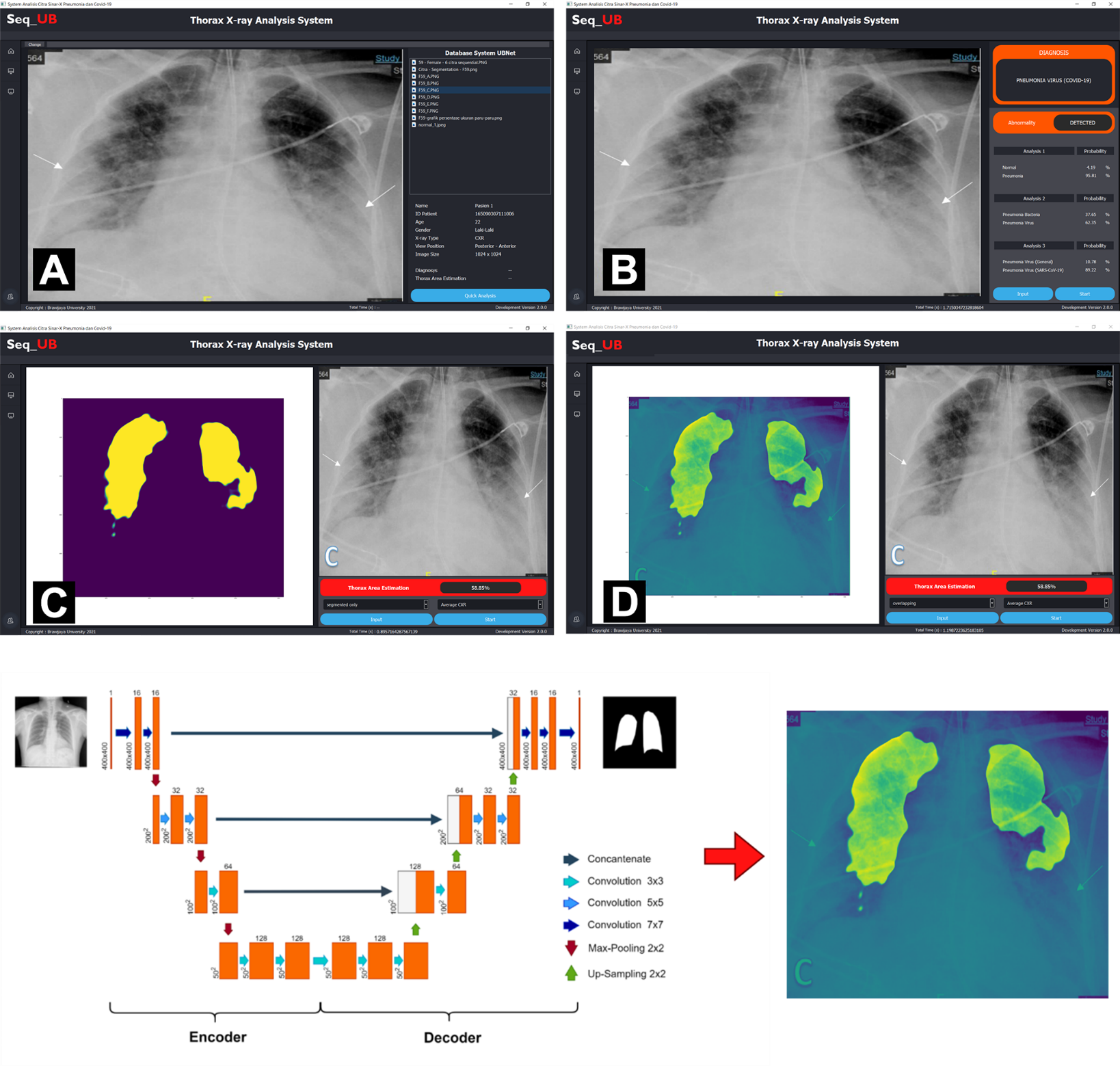
This research aims to develop an image segmentation algorithm, Seq_UB, which can monitor the development of the lung condition of Covid-19 patients periodically. The Seq_UB algorithm was developed by modifying the UNet system using different CNN layers according to the UBNet v1 algorithm.
Materials and methods
The dataset for training uses the Montgomery USA Dataset, which contains 138 CXRs with a resolution of 4,020×4,892, of which there are 80 normal CXR images and 58 CXR images of patients identified with tuberculosis; the Shenzhen Hospital Dataset consists of 662 CXR images, of which there are 336 abnormal CXR images.
Result and discussion
The results show that reducing the input image size does not significantly affect the accuracy of the segmentation results. However, reducing the input image size will affect the resolution of the segmentation results. Where the smaller the input image size, the lower the resolution obtained. This will have an impact on the final interpretation of the segmentation results. Research shows that an input image size of 512 is the best because the resolution of the segmentation results is still very accurate. This study shows that the Seq_UB architecture can perform X-ray image segmentation with relatively stable accuracy and lower computational burden. An interesting pattern was found, where Covid-19 patients quantitatively experienced fluctuations in image segmentation size.
Conclusions
The Seq_UB system can perform well with a segmentation accuracy of 96 %, and the processing speed takes 0.91 s. A desktop GUI was designed to segment X-ray images more effectively.
HIGHLIGHTS
- Development of the Seq_UB algorithm: This research presents the development of an X-ray image segmentation algorithm with a computational workload that is faster, more accurate, and lighter
- Quantitative estimation of the lung area percentage of Covid-19 patients based on segmentation with the Seg_UB model
- Monitoring the development of lung conditions of Covid-19 patients quantitatively by comparing it with normal CXR using the Seg_UB model
GRAPHICAL ABSTRACT
Downloads
References
K Ismail, H Bensasi, A Taha, A Nazir, M Abdelkhalek, W Mohamed, D Lodhe, S Buschbeck, M Bauer and Y Sakr. Characteristics and outcome of critically ill patients with coronavirus disease-2019 (covid-19) pneumonia admitted to a tertiary care center in the United Arab Emirates during the first wave of the SARS-CoV-2 pandemic. A retrospective analysis. PLos One 2021; 16, e0251687.
D Wootton and C Feldman. The diagnosis of pneumonia requires a chest radiograph (x-ray)-yes, no or sometimes? Pneumonia 2014; 5, 1-7.
G Besutti, PG Rossi, M Ottone, L Spaggiari, S Canovi, F Monelli, E Bonelli, T Fasano, N Sverzellati, A Caruso, N Facciolongo, G Ghidoni, A Simonazzi, M Iori, A Nitrosi, S Fugazzaro, S Costi, S Croci, E Teopompi, A Gallina, ..., C Salvarani. Inflammatory burden and persistent CT lung abnormalities in covid‑19 patients. Sci. Rep. 2022; 12, 4270.
S Waite, A Grigorian, RG Alexander, SL Macknik, M Carrasco, DJ Heeger and S Martinez-Conde. Analysis of perceptual expertise in radiology - current knowledge and a new perspective. Front. Hum. Neurosci. 2019; 13, 213.
YMY Abdallah and T Alqahtani. Research in medical imaging using image processing techniques. In: Y Zhou (Ed.). Medical Imaging. IntechOpen, London, 2019.
MAR Ahad, S Kobashi and JMRS Tavares. Advancements of image processing and vision in healthcare. J. Healthc. Eng. 2018; 2018, 8458024.
L Lu, X Wang, G Carneiro and L Yang. Deep learning and imaging and clinical networks for medical convolutional neural informatics. Advances in Computer Vision and Pattern Recognition. Springer Cham, Switzerland, 2019.
M Xin and Y Wang. Research on image classification model based on deep convolution neural network. EURASIP J. Image Video Process. 2019; 2019, 40.
S Srivastava, AV Divekar, C Anilkumar, I Naik, V Kulkarni and V Pattabiraman. Comparative analysis of deep learning image detection algorithms. J. Big Data 2021; 8, 66.
S Minaee, YY Boykov, F Porikli, AJ Plaza, N Kehtarnavaz and D Terzopoulos. Image segmentation using deep learning: A survey. IEEE Trans. Pattern Anal. Mach. Intell. 2021; 44, 3523-42.
A Aggarwal, M Mittal and G Battineni. Generative adversarial network: An overview of theory and applications. Int. J. Inform. Manag. Data Insights 2021; 1, 100004.
VV Danilov, D Litmanovich, A Proutski, A Kirpich, D Nefaridze, A Karpovsky and Y Gankin. Automatic scoring of covid-19 severity in x-ray imaging based on a novel deep learning workflow. Sci. Rep. 2022; 12, 12791.
O Ronneberger, P Fischer and T Brox. U-net: Convolutional networks for biomedical image segmentation. Lect. Notes Comput. Sci. 2015, https://doi.org/10.1007/978-3-319-24574-4_28.
CS Widodo, N Agus, MM Mahasin, Y Yueniwati, TA Putranto and PI Patra. UBNet: Deep learning-based approach for automatic x-ray image detection of pneumonia and covid-19 patients. J. X Ray Sci. Tech. 2021; 30, 57-71.
A Degerli, M Ahishali, M Yamac, S Kiranyaz, MEH Chowdhury, K Hameed, T Hamid, R Mazhar and M Gabbouj. Covid-19 infection map generation and detection from chest x-ray images. Health Inform. Sci. Syst. 2021; 9, 15.
S Jaeger, S Candemir, S Antani, YXJ Wáng, PX Lu and G Thoma. Two public chest x-ray datasets for computer-aided screening of pulmonary diseases. Quant. Imag. Med. Surg. 2014; 4, 475-7.
Y Tang, Y Tang, J Xiao and RM Summers. XLSor: A robust and accurate lung segmentor on chest x-rays using criss-cross attention and customized radiorealistic abnormalities generation. arXiv 2019, https://doi.org/10.48550/arXiv.1904.09229.
H Zhao and F Cao. Automatic lung segmentation algorithm on chest x-ray images based on fusion variational auto-encoder and three-terminal attention mechanism. Symmetry 2021; 13, 814.
E Shelhamer, J Long and T Darrell. Fully convolutional networks for semantic segmentation. IEEE Trans. Pattern Anal. Mach. Intell. 2015; 39, 640-51.
M Abadi, A Agarwal, P Barham, E Brevdo, Z Chen, C Citro, GS Corrado, A Davis, J Dean, M Devin, S Ghemawat, I Goodfellow, A Harp, G Irving, M Isard, Y Jia, R Jozefowicz, L Kaiser, M Kudlur, J Levenberg, ..., X Zheng. TensorFlow: Large-scale machine learning on heterogeneous systems. arXiv 2015, https://doi.org/10.48550/arXiv.1603.04467.
Keras, Available at: https://github.com/fchollet/keras, accessed March 2023.
R Yasin and W Gouda. Chest x-ray findings monitoring covid-19 disease course and severity. Egypt. J. Radiol. Nucl. Med. 2020; 51, 193.
HJ Chen, SJ Ruan, SW Huang and YT Peng. Lung x-ray segmentation using deep convolutional neural networks on contrast-enhanced binarized images. Mathematics 2020; 8, 545.
Z Zhou, MR Siddiquee, N Tajbakhsh and J Liang. UNet++: A nested u-net architecture for medical image segmentation. arXiv 2018, https://doi.org/10.48550/arXiv.1807.10165.
C Liang-Chieh, G Papandreou, F Schroff and H Adam. Rethinking atrous convolution for semantic image segmentation. arXiv 2017, https://doi.org/10.48550/arXiv.1706.05587.
C Liang-Chieh, Y Zhu, G Papandreou, F Schroff and H Adam. Encoder-decoder with atrous separable convolution for semantic image segmentation. arXiv 2018, https://doi.org/10.48550/arXiv.1802.02611.
A Kirillov and P Dollár. A unified architecture for instance and semantic segmentation object detection vs semantic segmentation. Universität Heidelberg, Germany, 2021.
A Chaurasia and E Culurciello. LinkNet: Exploiting encoder representations for efficient semantic segmentation. arXiv 2017, https://doi.org/10.1109/VCIP.2017.8305148.
H Zhao, J Shi, X Qi, X Wang and J Jia. Pyramid scene parsing network. In: Proceedings of the 2017 IEEE Conference on Computer Vision and Pattern Recognition, Hawaii. 2017.
H Li, P Xiong, J An and L Wang. Pyramid attention network for semantic segmentation. arXiv 2018, https://doi.org/10.48550/arXiv.1805.10180.
T Fan, G Wang, Y Li and H Wang. Ma-net: A multi-scale attention network for liver and tumor segmentation. IEEE Access 2020; 8, 179656-65.
Downloads
Published
How to Cite
Issue
Section
License
Copyright (c) 2023 Walailak University

This work is licensed under a Creative Commons Attribution-NonCommercial-NoDerivatives 4.0 International License.


