
The Potential of Sea Urchin (Diadema Setosum) Extracts as Antibacterial Against Staphylococcus Aureus
DOI:
https://doi.org/10.48048/tis.2024.7169Keywords:
Antibacterial, Fourier transform infrared, Sea urchin shell extract, Scanning electron microscope, Staphylococcus aureusAbstract
Secondary metabolites from sea urchin shells contain active substances with the potential of antibiotics. The purpose of this study was to investigate how sea urchin shell extract affected the development of the Staphylococcus bacterium. In this kind of research, the active chemical content of sea urchin shell extract was examined quantitatively, using scanning electron microscope (SEM) and Fourier Transform Infrared (FTIR), as well as the extract’s effect on the development of Staphylococcus aureus bacteria. According to the study, the average level of total flavonoids was 1.29 %, total alkaloids were 0.12 %, and total tannins were 1.0 %. Sea urchin shell powder has an absorbance of 0.42 at a wavelength of 1,400.84 cm–1. According to microbiological experiments, sea urchin shell ethyl acetate extract was able to stop Staphylococcus aureus growth in the category of the strong inhibition zone (inhibition zone diameter of 12 mm). Extract from sea urchin shells has antibacterial properties and may be used to make anti-diabetic ointments. This fabrication is a fantastic option for use in healthcare and medicine.
HIGHLIGHTS
- In this study, there are several main points including:
- The objective of this research is to determine the antibacterial activity of sea urchin shell extracts with various solvents.
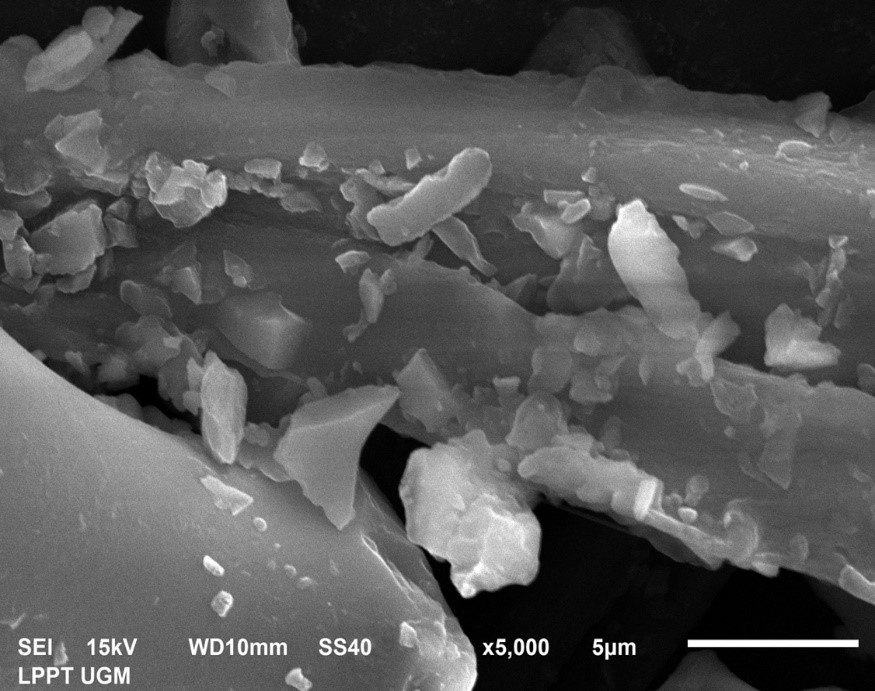
- FTIR and SEM results to determine the particle size of sea urchin shell powder and the compounds contained therein.
- Based on the results of the proximate test to see the antioxidant activity of the extract, it is known that the ethyl acetate extract contains the most flavonoid extract (1.6 %w/w).
- Ethyl acetate extract can inhibit the growth of Staphylococcus aureus bacteria in the Strong inhibition category.
GRAPHICAL ABSTRACT
Downloads
References
K Sapanli, T Kusumastanto, S Budiharsono and A Sadelie. Dinamika dan kebijakan pengembangan ekonomi kelautan Indonesia. Jurnal Kebijakan Sosial Ekonomi Kelautan Perikanan 2020; 10, 117-29.
NTB Gov. Propinsi Nusa Tenggara Barat, Available at: www.ntbprov.go.id, accessed May 2023.
Hafiluddin, Nurjanah and T Nurhayati. Kandungan gizi dan karakterisasi senyawa bioaktif lintah laut (Discodoris sp.). Jurnal Ilmiah Perikanan Kelautan 2011; 3, 1-6.
D Alwi, SH Muhammad and I Tae. Karakteristik morfologi dan indeks ekologi bulu babi (Echinoidea) di perairan desa wawama kabupaten pulau morotai. Jurnal Sumberdaya Akuatik Indopasifik 2020; 4, 23-32.
FR Arhas and S Kamal. Struktur komunitas dan karakteristik bulu babi (Echinoidea) di zona sublitoral perairan iboh kecamatan Sukakarya kota Sabang. In: Proceedings of the Prosiding Seminar Nasional Biotik, Aceh, Indonesia. 2018, p. 233-8.
E Yusron. Keanekaragaman jenis ekhinodermata di Perairan Likupang, Minahasa Utara, Sulawesi Utara. Ilmu Kelautan 2010; 15, 85-90.
SW Suwignyo, B Widigdo, YY Wardianto and M Krisanti. Avertebrata air. Penebar Swadaya, Jakarta, Indonesia, 2005.
AH Olii, MK Kadim, J Manajemen, S Perairan, F Perikanan and I Kelautan. Kepadatan dan pola sebaran bulu babi di desa lamu. Jurnal Ilmiah Perikanan Kelautan 2017; 5, 48-53.
LA Abubakar, CM Mwangi, JU Uku and SN Ndirangu. Antimicrobial activity of various extracts of the sea urchin Tripneustes gratilla (Echinoidea). Afr. J. Pharmacol. Therapeut. 2012; 1, 19-23.
A Kresnamurti, F Izazi and D Camelia. Standarisasi dan analisis FTIR ekstrak etanol 70 % bulu babi (Echinometra mathaei) dari sabang, nanggroe aceh darussalam standardization and FTIR analysis of 70 % ethanolic extract of Echinometra mathaei from sabang, nanggroe aceh darussalam. Farmasi 2022; 9, 1-8.
A Kresnamurti, F Izazi and D Kurniawati. Standardisasi ekstrak etanol 96 % bulu babi echinometra mathaei dari perairan bangkalan. J. Herbal Clin. Pharmaceut. Sci. 2021; 2, 21-8.
A Alavi, RG Sibbald, D Mayer, L Goodman, M Botros, DG Armstrong, K Woo, T Boeni, EA Ayello and RS Kirsner. Diabetic foot ulcers: Part 1. pathophysiology and prevention. J. Am. Acad. Dermatol. 2014; 70, 1.e1-1.e18.
P Chadwick, M Edmonds, J McCardle and D Armstrong. International Best practice guidelines: Wound management in diabetic foot ulcer. Wounds International, London, 2013.
Q Zhang, Y Wu and X Fei. Effect of probiotics on glucose metabolism in patients with type 2 diabetes mellitus: A meta-analysis of randomized controlled trials. Medicina 2016; 52, 28-34.
S Yusuf, M Okuwa, M Irwan, S Rassa, B Laitung, A Thalib, S Kasim, H Sanada, T Nakatani and J Sugama. Prevalence and risk factor of diabetic foot ulcers in a regional hospital, Eastern Indonesia. Open J. Nurs. 2016; 6, 1-10.
Y Vangoori, A Dakshinamoorthi and S Kavimani. Effect of myristica fragrans extract on lipid profile, glucose, body weight, food intake, liver and renal functions in experimental obese rats. Biomed. Pharmacol. J. 2019; 12, 677-82.
H Redel, Z Gao, H Li, AV Alekseyenko, Y Zhou, GI Perez-Perez, G Weinstock, E Sodergren and MJ Blaser. Quantitation and composition of cutaneous microbiota in diabetic and nondiabetic men. J. Infect. Dis. 2013; 207, 1105-14.
RD Desiati, E Sugiarti and S Ramandhany. Analisa ukuran partikel serbuk komposit nicral dengan penambahan reaktif elemen untuk aplikasi lapisan tahan panas. Metalurgi 2018; 1, 27-34.
HV Hoten. Analisis karakterisasi serbuk biokeramik dari cangkang telur ayam broiler. Jurnal Rotor 2020; 13, 1-5.
MI Darmawan, A Zaidah, A Hidayatulloh, IN Mentari, EF Utami and H Hardani. Performance of Cosmos caudatus chlorophyll dye on TiO2 nano particles coating in the manufacture of Dye-Sensitized Solar Cells (DSSC). J. Phys. Conf. Ser. 2021; 1869, 012108.
L Cui, HJ Butler, PL Martin-Hirsch and FL Martin. Aluminium foil as a potential substrate for ATR-FTIR, transflection FTIR or Raman spectrochemical analysis of biological specimens. Anal. Meth. 2016; 8, 481-7.
DE Méndez and T Allscher. Advantages of external reflection and transflection over ATR in the rapid material characterization of negatives and films via FTIR spectroscopy. Polymers 2022; 14, 808.
HM Si, Cari and A Supriyanto. Efficiency of dye-sensitized solar cell (DSSC) improvement as a light party TiO2-nano particle with extract pigment mangosteen peel (Garcinia mangostana). AIP Conf. Proc. 2014; 2014, 020002.
Hardani, S Idawati, BAA Mustariani, YK Dewi, A Hidayatulloh and MI Darmawan. Efficient TiO2 nanoparticle-ruthenium sensitizers with high open-circuit voltage (Voc) for high-performance dye-sensitized solar cells. J. Phys. Conf. Ser. 2021; 1816, 012005.
DR Adhika, AL Anindya, VV Tanuwijaya, H Rachmawati. Teknik pengamatan sampel biologi dan non-konduktif menggunakan. Scanning Electron Microsc. 2019, https://doi.org/10.5614/sniko.2018.9
ER Fischer, BT Hansen, V Nair, FH Hoyt and DW Dorward. Scanning electron microscopy. Curr. Protocol. Microbiol. 2012; 25, 1-47.
ER Fischer, BT Hansen, V Nair, FH Hoyt and DW Dorward. Nihms-375012, Available at: https://doi.org/10.1002/9780471729259.mc02b02s25.Scanning, accessed March 2023.
MD Murtey and P Ramasamy. Life science sample preparations for scanning electron microscopy. Acta Microscopica 2021; 30, 80-91.
H Hardani, MR Harahap and A Suhada. Ruthenium (N719) optimization to improve dye sensitized solar cell efficiency. Int. J. Thin Film Sci. Tech. 2022; 11, 47-53.
Y Tanaka, N Sasaki and A Ohmiya. Biosynthesis of plant pigments: Anthocyanins, betalains and carotenoids. Plant J. 2008; 54, 733-49.
NC Suryani, DGM Permana and AAGNA Jambe. Pengaruh jenis pelarut terhadap kandungan total flavonoid dan aktivitas aantioksidan ekstrak daun matoa (Pometia pinnata). Jurnal Ilmu Dan Teknologi Pangan 2016; 5, 1-10.
S Sudarmaji, Suhardi and B Haryono. Prosedur analisa untuk bahan makanan dan pertanian / oleh Slamet Sudarmaji, Bambang Haryono, Suhardi. Penerbit Liberty, Yogyakarta, Indonesian, 1984.
H Khotimah, Diyantoro, DW Indriati and AS Sundari. Screening in vitro antimicrobial activity of celery (Apium Graveolens) against Staphylococcus sp. Malays. J. Med. Health Sci. 2020; 16, 72-7.
RR Fadhilla, Diyantoro, DW Indriati and AS Sundari. Antibacterial potency of Indonesian randu honey against Staphylococcus sp. Malays. J. Med. Health Sci. 2020; 16, 67-71.
AS Sundari, DW Indriati and Diyantoro. Exploration of potential moraceae as an antimicrobial agent for coliform bacteria. Malays. J. Med. Health Sci. 2020; 16, 24-8.
AS Sundari, DW Indriati, Diyantoro, DW Indriati, H Ilmi, A Widyawaruyanti and AF Hafid. Screening of potential plants from kalimantan as an antimicrobial agent for coliform bacteria. Res. J. Pharm. Tech. 2022; 15, 4542-6.
WMM El-Sayed, MM Elshaer, HAH Ibrahim and MEA El-Metwaly. Antimicrobial agents from sea urchin (Diadema setosum) collected from the Red Sea, Egypt. Egypt. J. Aquat. Biol. Fish. 2020; 24, 33-51.
N Purwitasari, M Agil and H Studiawan. Activity of ethyl acetate fraction of merremia mammosa hall as anti-influenza a (H1N1). Indian J. Forensic Med. Toxicol. 2020; 14, 2070-3.
G Rompas, RAJ Lintang, DA Sumilat, IFM Rumengan, EL Ginting and H Pangkey. Aktivitas antibakteri dan analisis zookimia ekstrak bulu babi diadema setosum (Leske, 1778) asal perairan aertembaga, Kota Bitung. Jurnal Ilmiah PLATAX 2022; 10, 372-9.
H Hardani, DJ Sukmana, B Atfal and AD Pertiwi. Potential antimicrobial ethyl acetate extracts of ur burst shells against S.aureus bacteria from diabetic foot wounds. Jurnal Penelitian Pendidikan IPA 2023; 9, 1045-9.
A Apriandi, RMS Putri and I Tanjung. Karakterisasi, aktivitas antioksidan dan komponen bioaktif bulu babi (Diadema savignyi) dari perairan pantai trikora tiga pulau bintan. Majalah Ilmiah Biologi Biosfera 2020; 37, 49-54.
AD Pelu, R Tunny and B Latuconsina. Skrining fitokimia dan uji aktivitas antibakteri ekstrak etanol bulu babi (Diadema setosum) terhadap pertumbuhan staphylococcus aureus di perairan desa pelauw. Jurnal Sains Kesehatan 2020; 4, 1-15.
G Rompas, RAJ Lintang, DAA Sumilat, IFM Rumengan, EL Ginting and HD Pangkey. Antibacterial activity and zoochemical analysis of sea urchin Diadema setosum (Leske, 1778) extract from Aertembaga Waters, Bitung City. Jurnal Ilmiah Platax 2022; 10, 372.
A Amalia, I Sari and R Nursanty. Aktivitas antibakteri ekstrak etil asetat daun sembung (Blumea balsamifera (L.) DC.) terhadap pertumbuhan bakteri Methicillin Resistant Staphylococcus Aureus (MRSA). In: Proceedings of the Prosiding Seminar Nasional Biotik, Aceh, Indonesia. 2017, p. 387-91.
R Widowati, S Handayani and AR Al Fikri. Phytochemical screening and antibacterial activities of senggani (Melastoma malabathricum L.) ethanolic extract leaves. Jurnal Ilmu Pertanian Indonesia 2021; 26, 562-8.
I Rahmasari and ES Wahyuni. Efektivitas Memordoca carantia (pare) terhadap penurunan kadar glukosa darah. Jurnal Ilmiah Rekam Medis Informatika Kesehatan 2019; 9, 57-64.
R Khairunnisa, TU Soleha and MR Ramadhian. Identifikasi dan uji resistensi staphylococcus aureus pada ulkus diabetik di Instalasi Penyakit Dalam RSUD DR. H. Abdul Moeloek. J. Agromedicine Unila 2020; 7, 1-6.
YM Lee, YP Chong, M Kim, Y Eom, ES Kim, M Kim, KH Park, SH Kim, SO Lee, SH Choi, JH Woo and YS Kim. Long-term methicillin-resistant Staphylococcus aureus bacteremia persisting for more than 2 weeks: Risk factors and outcomes. Eur. J. Clin. Microbiol. Infect. Dis. 2019; 39, 773-81.

Downloads
Published
How to Cite
Issue
Section
License
Copyright (c) 2023 Walailak University

This work is licensed under a Creative Commons Attribution-NonCommercial-NoDerivatives 4.0 International License.


