
Investigation of Skin Discoloration in the Nile Tilapia (Oreochromis niloticus) by Plasma-Activated Media
DOI:
https://doi.org/10.48048/tis.2026.13971Keywords:
Nile tilapia, Cold plasma, Plasma-activated media, Melanophore density, Growth performance, Reactive oxygen and nitrogen species (RONS), Skin pigmentation, Nile tilapia, Cold plasma, Plasma-activated media, Melanophore density, Growth performance, Reactive oxygen and nitrogen species (RONS), Skin pigmentationAbstract
This research investigated the efficacy of plasma-activated media (PAM) as a tool for inducing stable skin color variants in Nile tilapia (Oreochromis niloticus). PAM was generated using a transient corona discharge system (20 W,
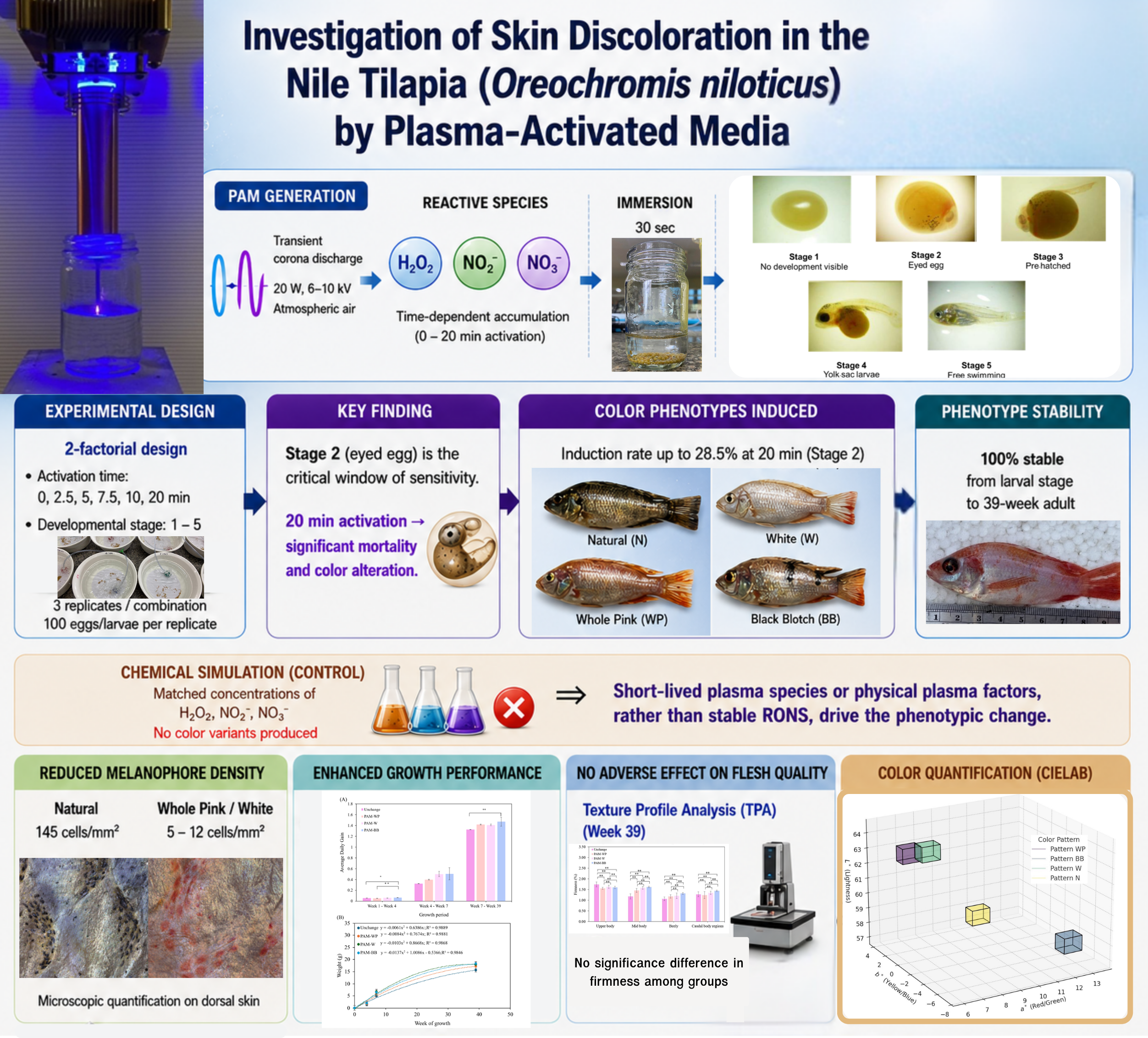
6 - 10 kV) in atmospheric air, resulting in the time-dependent accumulation of hydrogen peroxide, nitrite, and nitrate. A 2-factorial experimental design was employed to evaluate the interaction between PAM activation duration (0–20 min) and developmental stage (Stages 1 - 5). Survival analysis at 14 days post-immersion identified Stage 2 (eyed egg) as a critical window of sensitivity, showing significant mortality and phenotypic shifts at the 20-minute activation level. In contrast, Stage 3 (pre-hatched embryo) and later larval stages demonstrated high tolerance to PAM exposure. PAM immersion successfully induced 4 distinct color phenotypes: Natural, Whole Pink, White, and Black Blotch, with an induction rate of 28.5% in the most intense treatment group. Notably, a chemically simulated control group matching the PAM’s RONS concentrations failed to produce any variants, suggesting that short-lived species or physical plasma factors are the primary drivers of the phenotypic shift. The induced phenotypes were 100% stable from the larval stage through the 39-week adult harvest. Quantitative CIELAB analysis and microscopic quantification revealed that Whole Pink and White phenotypes were characterized by a significant reduction in melanophore density (5 - 12 cells/mm2) compared to the Natural control (145 cells/mm2). Long-term monitoring demonstrated that PAM-treated groups exhibited significantly higher body weights and Average Daily Gain (ADG) by Week 39, consistent with a hermetic response. Texture Profile Analysis (TPA) confirmed that PAM exposure did not negatively affect flesh firmness across anatomical regions. These results indicate that PAM immersion is a safe, stable, and non-genetic method for inducing commercially desirable color variants and enhancing growth performance in Nile tilapia.
HIGHLIGHTS
- Plasma-activated media induced stable skin color variants in Nile tilapia.
- Stage 2 eyed eggs were the most sensitive window for color induction.
- Whole Pink and White phenotypes remained stable until 39 weeks of growth.
- PAM treatment enhanced growth performance without reducing flesh firmness.
- Short-lived plasma species likely contributed to pigmentation changes.
GRAPHICAL ABSTRACT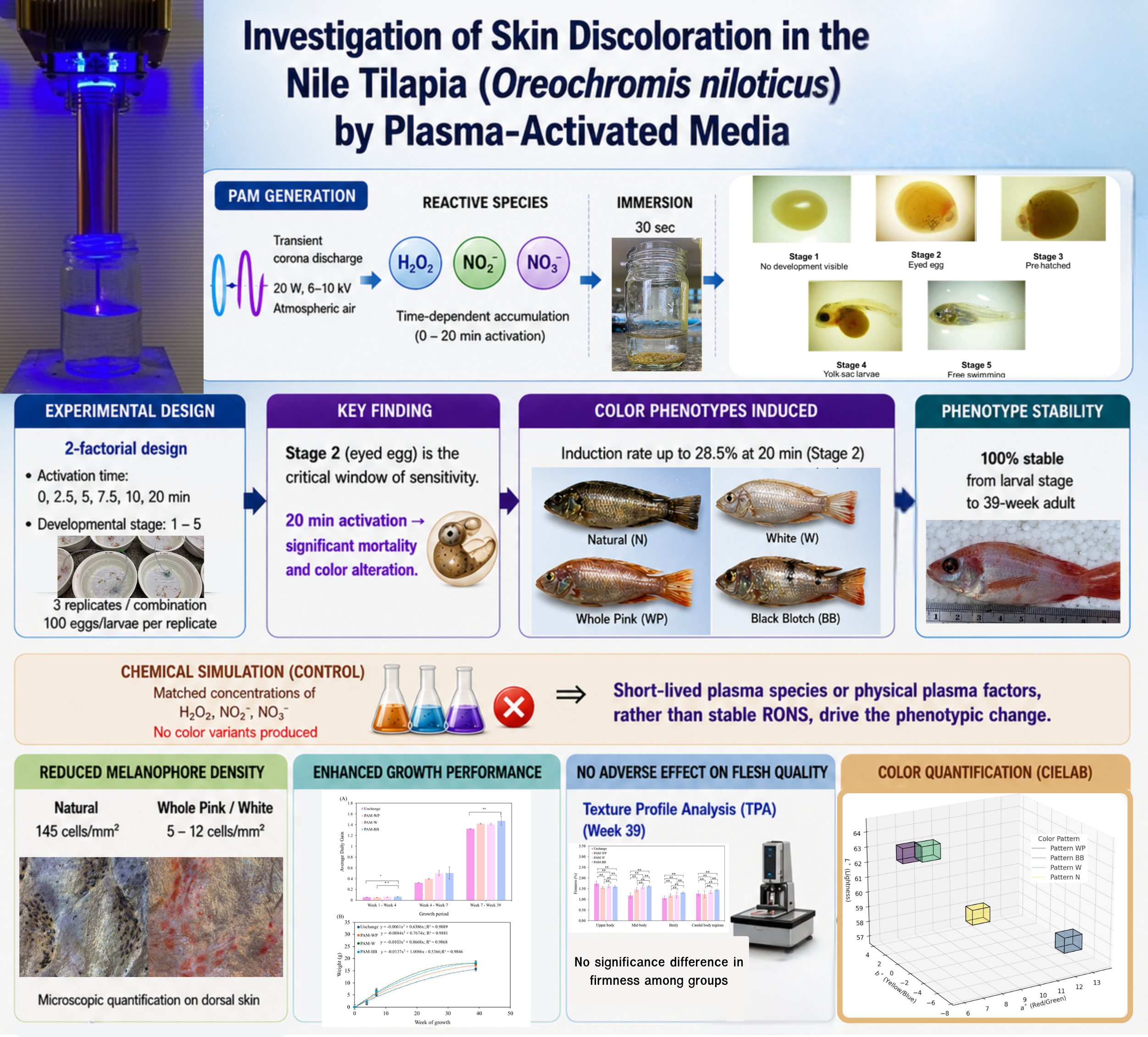
Downloads
References
ME Abd El-Hack, MT El-Saadony, MM Nader, HM Salem, AM El-Tahan, SM Soliman and AF Khafaga. Effect of environmental factors on growth performance of Nile tilapia (Oreochromis niloticus). International Journal of Biometeorology 2022; 66(11), 2183-2194.
O Nwachi, Y Esa, A Christianus, A Rahim and M Kamarudin. Patterns of colour inheritance from crossbreeding between Red hybrid tilapia (Oreochromis sp.) and GIFT tilapia (Oreochromis niloticus). Journal of Environmental Biology 2020; 41, 1289-1294.
M Luo, G Lu, H Yin, L Wang, M Atuganile and Z Dong. Fish pigmentation and coloration: Molecular mechanisms and aquaculture perspectives. Reviews in Aquaculture 2021; 13(4), 2395-2412.
J Liu, M Yin, Z Ye, J Hu and Z Bao. Harnessing hue: Advances and applications of fish skin pigmentation genetics in aquaculture. Fishes 2024; 9(6), 220.
M Domonkos, P Tichá, J Trejbal and P Demo. Applications of cold atmospheric pressure plasma technology in medicine, agriculture and food industry. Applied Sciences 2021; 11(11), 4809.
W Nupangtha, C Kuensaen, S Sarapirom and D Boonyawan. Investigation of Cold Atmospheric Plasma induce Apoptosis in Cancer Cell. IOP Conference Series: Journal of Physics: Conference Series 2018; 1144(1), 012025.
P Poramapijitwat, P Thana, D Boonyawan, K Janpong, C Kuensaen, W Charerntantanakul, LD Yu and S Sarapirom. Effect of dielectric barrier discharge plasma jet on bactericidal and human dermal fibroblasts adult cells: In vitro contaminated wound healing model. Surface and Coatings Technology 2020; 402, 126482.
D Boonyawan, K Lamasai, C Umongno, S Rattanatabtimtong, LD Yu, C Kuensaen, J Maitip and P Thana. Surface dielectric barrier discharge plasma-treated pork cut parts: bactericidal efficacy and physiochemical characteristics. Heliyon 2022; 8(10), e10915.
P Thana, D Boonyawan, M Jaikua, W Promsart, A Rueangwong, S Ungwiwatkul, K Prasertboonyai and J Maitip. Plasma-activated water (PAW) decontamination of foodborne bacteria in shucked oyster meats using a compact flow-through generator. Foods 2025; 14(9), 1502.
K Moonsub, P Seesuriyachan, D Boonyawan, P Rachtanapun, C Sawangrat, T Opassuwan and W Wattanutchariya. Combating foodborne pathogens: Efficacy of plasma-activated water with supplementary methods for Staphylococcus aureus eradication on chicken, and beef. Food Chemistry: X 2024; 24, 101890.
K Kosumsupamala, P Thana, N Palee, K Lamasai, C Kuensaen, A Ngamjarurojana, P Yangkhamman and D Boonyawan. Air to H2-N2 pulse plasma jet for in-vitro plant tissue culture process: source characteristics. Plasma Chemistry and Plasma Processing 2022; 42(3), 535-559.
A Sakulthai, C Sawangrat, D Pichpol, J Kongkapan, T Srikanchai, R Charoensook, P Sojithamporn and D Boonyawan. Improving the efficiency of crossbred Pradu Hang Dam chicken production for meat consumption using cold plasma technology on eggs. Scientific Reports 2023; 13(1), 2836.
Y Ikeda, R Fukushima, K Tange, H Motomura, T Saito and M Jinno. Growth acceleration of Nile tilapia at 21 to 31 weeks of age with plasma-treated air-supplied water. Free Radical Research 2023; 57(1), 21-29.
A Waskow, A Howling and I Furno. Mechanisms of plasma-seed treatments as a potential seed processing technology. Frontiers in Physics 2021; 9, 617345.
N Puač, M Gherardi and M Shiratani. Plasma agriculture: A rapidly emerging field. Plasma Processes and Polymers 2018; 15(2), 1700174.
DM Parichy. Evolution of pigment cells and patterns: Recent insights from teleost fishes. Current Opinion in Genetics & Development 2021; 69, 88-96.
L Wang, B Jiang, W Zhu, J Fu, M Luo, W Liu and Z Dong. The role of melanocortin 1 receptor on melanogenesis pathway in skin color differentiation of red tilapia. Aquaculture Reports 2022; 22, 100946
JA Lister. Development of pigment cells in the zebrafish embryo. Microscopy Research and Technique 2002; 58(6), 435-441.
TY Ma, JY Wu, XK Gao, JY Wang, XL Zhan and WS Li. Molecular cloning, functional identification and expressional analyses of FasL in Tilapia, Oreochromis niloticus. Developmental & Comparative Immunology 2014; 46(2), 448-460.
A Sharifian, N Soltanizadeh and R Abbaszadeh. Effects of dielectric barrier discharge plasma on the physicochemical and functional properties of myofibrillar proteins. Innovative Food Science & Emerging Technologies 2019; 54, 1-8.
K Fujimura and N Okada. Development of the embryo, larva and early juvenile of Nile tilapia Oreochromis niloticus (Pisces: Cichlidae). Developmental staging system. Development, Growth & Differentiation 2007; 49(4), 301-324.
EM Konchekov, N Gusein-zade, DE Burmistrov, LV Kolik, AS Dorokhov, AY Izmailov, B Shokri and SV Gudkov. Advancements in plasma agriculture: A review of recent studies. International Journal of Molecular Sciences 2023; 24(20), 15093.
G Kwikiriza, I Abaho, PD Tibihika, AA Izaara, F Atukwatse, T Omara, JK Nattabi, N Kasozi, M Curto and H Meimberg. Genetic diversity and population differentiation of farmed nile tilapia (Oreochromis niloticus Linnaeus, 1758) to advance selective breeding in Uganda. Diversity 2025; 17(2), 128.
DM Parichy and JE Spiewak. Origins of adult pigmentation: diversity in pigment stem cell lineages and implications for pattern evolution. Pigment Cell & Melanoma Research 2015; 28(1), 31-50.
L Wang, W Zhu, Z Dong, F Song, J Dong and J Fu. Comparative microRNA-seq analysis depicts candidate miRNAs involved in skin color differentiation in red tilapia. International Journal of Molecular Sciences 2018; 19(4), 1209.
L Reich, J Don and R Avtalion. Inheritance of the red color in tilapias. Genetica 1990; 80(3), 195-200.
B Jiang, L Wang, M Luo, J Fu, W Zhu, W Liu and Z Dong. Transcriptome analysis of skin color variation during and after overwintering of Malaysian red tilapia. Fish Physiology and Biochemistry 2022; 48(3), 669-682.
F Song, L Shi, F Yao, Y Gu, D Zheng, W Zhang, Y Liang, K Zhang, M Yang and J Luo. The effect of background color on skin color variation of juvenile Plectropomus leopardus. Animals 2022; 12(23), 3349.
M Hamed, AG Osman, AE Badrey, HA Soliman and AEDH Sayed. Microplastics-induced eryptosis and poikilocytosis in early-juvenile nile Tilapia (Oreochromis niloticus). Frontiers in Physiology 2021; 12, 742922.
KS Wong, NS Chew, M Low and MK Tan. Plasma-activated water: Physicochemical properties, generation techniques, and applications. Processes 2023; 11(7), 2213.
O Olagunju. 2008, Peroxynitrite chemistry: Formation, decomposition and possible deactivation mechanisms by thiols. Ph. D. Dissertation. Portland State University, Portland, United States, 2008.
A Phaniendra, DB Jestadi and L Periyasamy. Free radicals: Properties, sources, targets, and their implication in various diseases. Indian Journal of Clinical Biochemistry 2015; 30(1), 11-26.
ADG Sampaio, W Chiappim, NVM Milhan, BB Neto, R Pessoa and CY Koga-Ito. Effect of the pH on the antibacterial potential and cytotoxicity of different plasma-activated liquids. International Journal of Molecular Sciences 2022; 23(22), 13893.
P Lukes, E Dolezalova, I Sisrova and M Clupek. Aqueous-phase chemistry and bactericidal effects from an air discharge plasma in contact with water: Evidence for the formation of peroxynitrite through a pseudo-second-order post-discharge reaction of H2O2 and HNO2. Plasma Sources Science and Technology 2014; 23(1), 015019.
PR Rotondo, D Aceto, M Ambrico, AM Stellacci, F Faretra, RMD Angelini and PF Ambrico. Physicochemical properties of plasma-activated water and associated antimicrobial activity against fungi and bacteria. Scientific Reports 2025; 15(1), 5536.
S Gligorovski, R Strekowski, S Barbati and D Vione. Environmental implications of hydroxyl radicals (•OH). Chemical Reviews 2015; 115(24), 13051-13092.
M Naïtali, JM Herry, E Hnatiuc, G Kamgang and JL Brisset. Kinetics and bacterial inactivation induced by peroxynitrite in electric discharges in air. Plasma Chemistry and Plasma Processing 2012; 32(4), 675-692.
S Kalghatgi, CM Kelly, E Cerchar, B Torabi, O Alekseev, A Fridman, G Friedman and J Azizkhan-Clifford. Effects of non-thermal plasma on mammalian cells. PLoS One 2011; 6(1), e16270.
JL Duband. Neural crest delamination and migration. In: JP Saint-Jeannet (Ed.). Neural crest induction and differentiation. advances in experimental medicine and biology. Springer, Boston, United States, 2006, p. 45-77.
JD Berndt, MR Clay, T Langenberg and MC Halloran. Rho-kinase and myosin II affect dynamic neural crest cell behaviors during epithelial to mesenchymal transition in vivo. Developmental Biology 2008; 324(2), 236-244.
LM San-Jose and A Roulin. On the potential role of the neural crest cells in integrating pigmentation into behavioral and physiological syndromes. Frontiers in Ecology and Evolution 2020; 8, 278.
F Yu, Y Lu, Z Zhong, B Qu, M Wang, X Yu and J Chen. Mitf involved in innate immunity by activating tyrosinase-mediated melanin synthesis in Pteria penguin. Frontiers in Immunology 2021; 12, 626493.
T Hida, T Kamiya, A Kawakami, J Ogino, H Sohma, H Uhara and K Jimbow. Elucidation of melanogenesis cascade for identifying pathophysiology and therapeutic approach of pigmentary disorders and melanoma. International Journal of Molecular Sciences 2020; 21(17), 6129.
R Sauermann, B Fancourt, T Faulkner, H Shute, D Reid, AJ Pask and CY Feigin. Loss-of-function mutations in ASIP and MC1R are associated with coat colour variation in marsupials. Biology Letters 2025; 21(10), 20250302.
UW Khan, Ø Øverli, PM Hinkle, FA Pasha, IB Johansen, I Berget, PIM Silva, S Kittilsen, E Höglund, SW Omholt and DI Våge. A novel role for pigment genes in the stress response in rainbow trout (Oncorhynchus mykiss). Scientific Reports 2016; 6(1), 28969.
Z Qi, J Liu, J Shi, M Yin, J Liu, J Fan, Z Bao, Z Ye and J Hu. Integrated transcriptomic and epigenomic analysis reveals mechanisms underlying melanotic spot formation in red tilapia (Oreochromis spp.). International Journal of Molecular Sciences 2025; 26(9), 4370.
J Pérez-Sánchez, P Simó-Mirabet, F Naya-Català, JA Martos-Sitcha, E Perera, A Bermejo-Nogales, L Benedito-Palos and JA Calduch-Giner. Somatotropic axis regulation unravels the differential effects of nutritional and environmental factors in growth performance of marine farmed fishes. Frontiers in Endocrinology 2018; 9, 687.
Y Song, S Chen, L Li, Y Zeng and X Hu. The hypopigmentation mechanism of tyrosinase inhibitory peptides derived from food proteins: An overview. Molecules 2022; 27(9), 2710.
ET Won and RJ Borski. Endocrine regulation of compensatory growth in fish. Frontiers in Endocrinology 2013; 4, 74.
T Nakano, LO Afonso, BR Beckman, GK Iwama and RH Devlin. Acute physiological stress down-regulates mRNA expressions of growth-related genes in coho salmon. PLoS One 2013; 8(8), e71421.
HP Chen, L Zhang, BH Xu, LM Kang, SL Yang, HH Zeng and XY Shi. Nitric oxide stimulates embryonic somatotroph differentiation and growth hormone mRNA and protein expression through a cyclic guanosine monophosphate-independent mechanism. Tissue and Cell 2009; 41(2), 133-140.
Z Li, P Meekuan, YX Wang, ZH Feng, SY Luo, ZX Zhang, J Xiao, F Yu and ZS Hou. Effects of Different Salinity Conditions on Regulation of ghrh-sst-gh-igf Axis in Nile Tilapia (Oreochromis niloticus): Insights from Transcriptional Signature. International Journal of Molecular Sciences 2025; 26(17), 8261.
PG Vissio, MJ Darias, MP Di Yorio, DIP Sirkin and TH Delgadin. Fish skin pigmentation in aquaculture: The influence of rearing conditions and its neuroendocrine regulation. General and Comparative Endocrinology 2021; 301, 113662.
OO Olatunde, A Singh, KA Shiekh, P Nuthong and S Benjakul. Effect of high voltage cold plasma on oxidation, physiochemical, and gelling properties of myofibrillar protein isolate from Asian sea bass (Lates calcarifer). Foods 2021; 10(2), 326.
S Rout, PK Panda, P Dash, PP Srivastav and CT Hsieh. Cold plasma-induced modulation of protein and lipid macromolecules: A review. International Journal of Molecular Sciences 2025; 26(4), 1564.
SK Pankaj, Z Wan and KM Keener. Effects of cold plasma on food quality: A review. Foods 2018; 7(1), 4.
J Jirešová, V Scholtz, J Julák and B Šerá. Comparison of the effect of plasma-activated water and artificially prepared plasma-activated water on wheat grain properties. Plants 2022; 11(11), 1471.
T Yang, W Deng, L Liu, G Xia, L Zhang and J Wang. Effects of plasma-activated lactic acid on the physicochemical properties and volatile organic compounds of refrigerated tilapia fillets. Food Chemistry: X 2026; 34, 103491.
Downloads
Published
How to Cite
Issue
Section
License
Copyright (c) 2026 Walailak University

This work is licensed under a Creative Commons Attribution-NonCommercial-NoDerivatives 4.0 International License.