
Fabrication and Cytotoxicity Testing of Silver Nitrate Modified PETG Scaffolds for Bone Applications
DOI:
https://doi.org/10.48048/tis.2026.12373Keywords:
PETG, silver nitrate (AgNO3), Coating, 3D printing, Surface modification, Laydown patterns, Bone tissue applications, CytocompatibilityAbstract
This study examines the surface modification of 3D printed polyethylene terephthalate glycol (PETG) scaffolds by a simple and reproducible coating method using silver nitrate (AgNO3) and the cytotoxicity of the modified scaffolds is assessed. The main aims of the study were characterization of the structural and morphological changes on the surface of the scaffold after the treatment, more precisely the thickness of the deposited silver coating, and assessment of the biological effect on the HBL100 normal cell lines. The chemical protocol for scaffold modification was a multi-step process including the preparation of silver nitrate solution, a necessary pre-treatment of PETG scaffold using a sodium hydroxide (NaOH) solution, immersion of scaffold in the AgNO3 solution, sonication and final drying process. Atomic Force Microscopy (AFM) analysis was performed to measure the thickness of the coating, finding a high dependence of the coating on the 3D printing angle of the scaffold. The PETG scaffold with 0°/45° had a coating thickness of about 566 nm, which was much thicker than coatings on scaffolds with 0°/60°/120° and 0°/90° printing laydown patterns. The cytotoxicity of the scaffolds was tested by the standard MTT assay. The results of the assay including mean absorbance, mean inhibition percentage and standard deviation showed no significant cytotoxic effect from the coated scaffolds with the mean inhibition percentages ranging from a minimal 0.74% to 10.91%. This study demonstrates a viable and biocompatible method to functionalize 3D-printed PETG scaffolds which could have significant implications for various applications including tissue engineering and medical devices where the need to both structural integrity and non-toxic surface properties is paramount.
HIGHLIGHTS
- The laydown pattern of 3D printing has a substantial impact on the thickness of silver nitrate coating on PETG scaffolds, varying from 89 nm (0°/60°/120°) to 566 nm (0°/45°) that allows rational surface functionalization.
- It is demonstrated that NaOH pretreatment and AgNO3 immersion is a simple and reproducible chemical coating method for functionalization of 3D printed PETG scaffolds with antimicrobial capabilities without significant structural degradation.
- Silver-coated PETG scaffolds have shown great biocompatibility with normal HBL100 cell lines with low cytotoxicity (0.74% - 10.91% inhibition) for all different printing geometries.
- Characterization AFM The geometry-dependent surface morphology of 0°/45° scaffolds reveal thick and rough coatings with aggregation of nanoparticles whereas and 0°/90° scaffolds reveal thin and uniform coatings ideal towards mechanical stability.
- Multifunctional bone scaffolds by combining the mechanical properties of PETG with the antimicrobial activity of silver have promising potential for use in bone regeneration applications where infection is a common problem without any decline in cellular compatibility.
GRAPHICAL ABSTRACT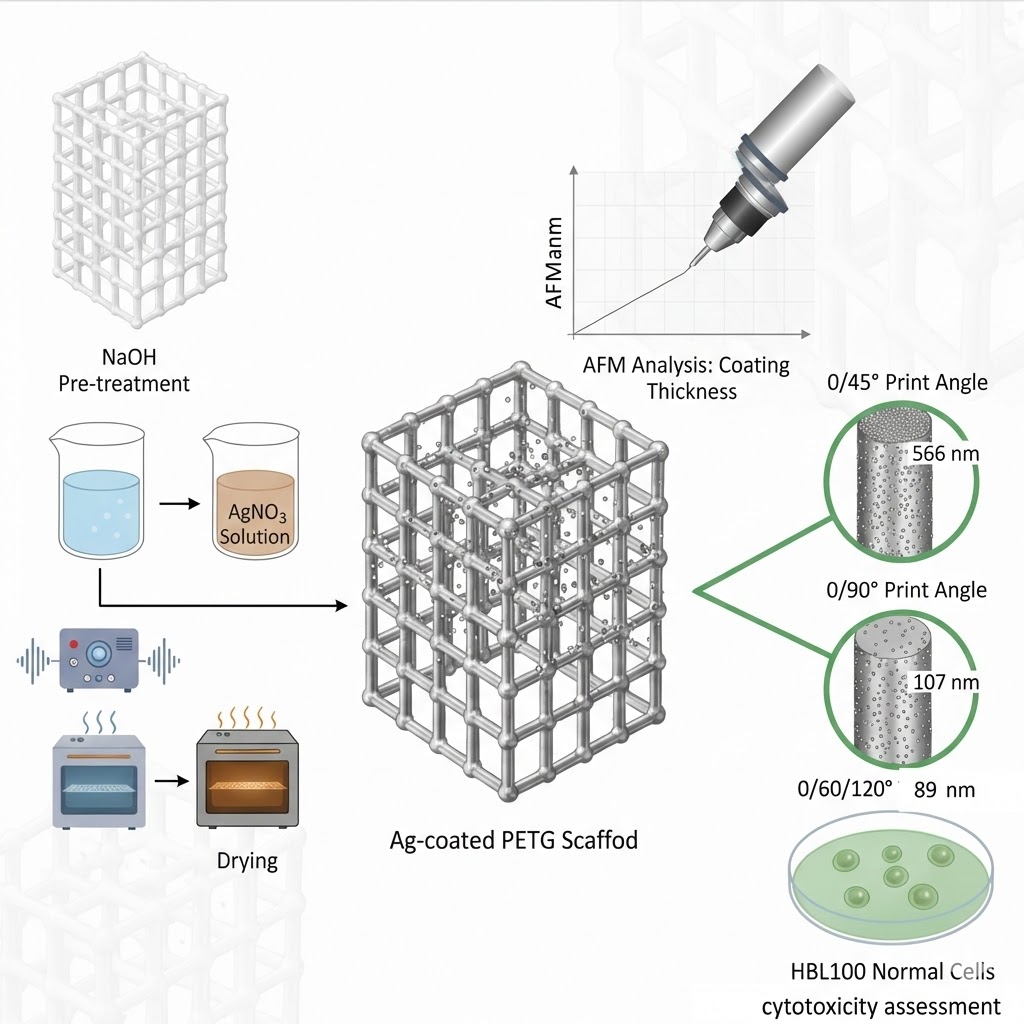
Downloads
References
J Meneses, JC Silva, SR Fernandes, A Datta, F Castelo Ferreira, C Moura, S Amado, N Alves and P Pascoal-Faria. A multimodal stimulation cell culture bioreactor for tissue engineering: A numerical modelling approach. Polymers 2020; 12(4), 940.
G Iosub, IA Lungescu, AC Bîrcă, AG Niculescu, PC Balaure, S Constantinescu, B Mihaiescu, DM Rădulescu, AM Grumezescu, A Hudiță, IA Neacșu and AR Rădulescu. New three dimensional-printed polyethylene terephthalate glycol liners for hip joint endoprostheses: A bioactive platform for bone regeneration. Materials 2025; 18(6), 1206.
MH Hassan, AM Omar, E Daskalakis, Y Hou, B Huang, I Strashnov, BD Grieve and P Bártolo. The potential of polyethylene terephthalate glycol as biomaterial for bone tissue engineering. Polymers 2020; 12(12), 3045.
J Sun, H Zhu, H Wang, J Li, B Li, L Liu and H Yang. A multifunctional composite scaffold responds to microenvironment and guides osteogenesis for the repair of infected bone defects. Journal of Nanobiotechnology 2024; 22(1), 577.
L Qin, S Yang, C Zhao, J Yang, F Li, Z Xu, Y Yang, H Zhou, K Li, C Xiong, W Huang, N Hu and X Hu. Prospects and challenges for the application of tissue engineering technologies in the treatment of bone infections. Bone Research 2024; 12(1), 28.
JCC Paiva, L Oliveira, MF Vaz and S Costa-de-Oliveira. Biodegradable bone implants as a new hope to reduce device-associated infections - a systematic review. Bioengineering 2022; 9(8), 409.
JCC Paiva, L Oliveira, MF Vaz and S Costa-de-Oliveira. Construction of antibacterial bone implants and their application in bone regeneration. Materials Horizons 2024; 11(3), 590-625.
CT Johnson and AJ García. Scaffold-based anti-infection strategies in bone repair. Annals of Biomedical Engineering 2015; 43, 515-528.
M Li, P Zhao, J Wang, X Zhang and J Li. Functional antimicrobial peptide-loaded 3D scaffolds for infected bone defect treatment with AI and multidimensional printing. Materials Horizons 2025; 12(1), 20-36.
A Roy, P Basuthakur, S Haque and CR Patra. Silver‐based nanoparticles for antibacterial activity: Recent development and mechanistic approaches. In: RN Krishnaraj and RK Sani (Eds.). Microbial interactions at nanobiotechnology interfaces: Molecular mechanisms and applications. Wiley, New Jersey, 2021, p. 245-301.
M Mohiti-Asli, B Pourdeyhimi and EG Loboa. Novel, silver-ion-releasing nanofibrous scaffolds exhibit excellent antibacterial efficacy without the use of silver nanoparticles. Acta Biomaterialia 2014; 10(5), 2096-2104.
SS Goderecci. 2016, Cytotoxic and antimicrobial effects of silver-containing surfaces. Master Thesis. Rowan University, New Jersey, 2016.
L Xu, YY Wang, J Huang, CY Chen, ZX Wang and H Xie. Silver nanoparticles: Synthesis, medical applications and biosafety. Theranostics 2020; 10(20), 8996.
E Hamidi. Antibacterial coating for aesthetic orthodontic devices, Available at: https://www.politesi.polimi.it/handle/10589/187701, accessed July 2025.
D Flores, J Noboa, M Tarapues, K Vizuete, A Debut, L Bejarano, DA Streitwieser and S Ponce. Simple preparation of metal-impregnated FDM 3D-printed structures. Micromachines 2022; 13(10), 1675.
X Wu, J Li, L Wang, D Huang, Y Zuo and Y Li. The release properties of silver ions from Ag-nHA/TiO2/PA66 antimicrobial composite scaffolds. Biomedical Materials 2010; 5(4), 44105.
R Zhu, X Li, C Wu, L Du, X Du and T Tafsirojjaman. Effect of hydrothermal environment on mechanical properties and electrical response behavior of continuous Carbon Fiber/Epoxy composite plates. Polymers 2022; 14(19), 4072.
H Qin, C Zhu, Z An, Y Jiang, Y Zhao, J Wang, X Liu, B Hui, X Zhang and Y Wang. Silver nanoparticles promote osteogenic differentiation of human urine-derived stem cells at noncytotoxic concentrations. International Journal of Nanomedicine 2014; 4, 2469-2478.
L Sethuram, J Thomas, A Mukherjee and N Chandrasekaran. Effects and formulation of silver nanoscaffolds on cytotoxicity dependent ion release kinetics towards enhanced excision wound healing patterns in Wistar albino rats. RSC Advances 2019; 9(61), 35677-35694.
A Sati, TN Ranade, SN Mali, HKA Yasin and A Pratap. Silver nanoparticles (AgNPs): Comprehensive insights into bio/synthesis, key influencing factors, multifaceted applications, and toxicity - a 2024 update. ACS Omega 2025; 10(8), 7549-7582.
S Maruelli, R Besio, J Rousseau, N Garibaldi, J Amiaud, B Brulin, P Layrolle, V Escriou and A Forlino. Osteoblasts mineralization and collagen matrix are conserved upon specific Col1a2 silencing. Matrix Biology Plus 2020; 6, 100028.
MG Kontakis, E Carlsson, C Palo-Nieto and NP Hailer. Ionic silver coating of orthopedic implants may impair osteogenic differentiation and mineralization. Experimental and Therapeutic Medicine 2025; 29(3), 51.
Y Xu, B Zheng, J He, Z Cui and Y Liu. Silver nanoparticles promote osteogenic differentiation of human periodontal ligament fibroblasts by regulating the RhoA-TAZ axis. Cell Biology International 2019; 43(8), 910-920.
E Dube and GE Okuthe. Silver nanoparticle-based antimicrobial coatings: Sustainable strategies for microbial contamination control. Microbiology Research 2025; 16(6), 110.
T Liu, G Yang, T Li, Q Wang, H Liu and F He. Preparation of Ag@ 3D‐TiO2 scaffolds and determination of its antimicrobial properties and osteogenesis‐p romoting ability. Orthopaedic Surgery 2024; 16(6), 1445-1460.
SK Nemani, RK Annavarapu, B Mohammadian, A Raiyan, J Heil, A Haque, A Abdelaal and H Sojoudi. Surface modification of polymers: Methods and applications. Advanced Materials Interfaces 2018; 5(24), 1801247.
R Subramani, RR Leon, R Nageswaren, MA Rusho and KV Shankar. Tribological performance enhancement in FDM and SLA additive manufacturing: Materials, mechanisms, surface engineering, and hybrid strategies - a holistic review. Lubricants 2025; 13(7), 298.
AG Khina and YA Krutyakov. Similarities and differences in the mechanism of antibacterial action of silver ions and nanoparticles. Applied Biochemistry and Microbiology 2021; 57(6), 683-693.
A Merlo, E González-Martínez, K Saad, M Gomez, M Grewal, J Deering, LA DiCecco, Z Hosseinidoust, KN Sask, JM Moran-Mirabal and K Grandfield. Functionalization of 3d printed scaffolds using polydopamine and silver nanoparticles for bone-interfacing applications. ACS Applied Bio Materials 2023; 6(3), 1161-1172.
NK Dhiman, S Agnihotri and R Shukla. Silver-based polymeric nanocomposites as antimicrobial coatings for biomedical applications. In: S Singh and P Maurya (Eds.). Nanotechnology in modern animal biotechnology: Recent trends and future perspectives. Springer, Singapore, 2019, p. 115-171.
H Shao, T Zhang, Y Gong and Y He. Silver‐containing biomaterials for biomedical hard tissue implants. Advanced Healthcare Materials 2023; 12(26), 2300932.
M Harun-Ur-Rashid, T Foyez, SBN Krishna, S Poda and AB Imran. Recent advances of silver nanoparticle-based polymer nanocomposites for biomedical applications. RSC Advances 2025; 15(11), 8480-8505.
Y Huang, Y Zhang, J Sheng, Z Li, W Zhang, J Shen, S Yang, J Zhong, L Yu and X Chen. Study on the preparation and properties of 3D‐printed PETG/AgNPs antibacterial coatings for clear aligners. Polymer Composites 2025; 46(17), 15849-15860.
C Yan, C Kleiner, A Tabigue, V Shah, G Sacks, D Shah and V DeStefano. PETG: Applications in modern medicine. Engineered Regeneration 2024; 5(1), 45-55.
ME Astaneh and N Fereydouni. Silver nanoparticles in 3D printing: A new frontier in wound healing. ACS Omega 2024; 9(40), 41107-41129.
MUA Khan, SIA Razak, H Mehboob, MRA Kadir, TJS Anand, F Inam, SA Shah, MEF Abdel-Haliem and R Amin. Synthesis and characterization of silver-coated polymeric scaffolds for bone tissue engineering: antibacterial and in vitro evaluation of cytotoxicity and biocompatibility. ACS Omega 2021; 6(6), 4335-4346.
H Mishbak, MH Hassan, E Daskalakis, AM Omar, DM Freitas, W Mirihanage, P Mativenga, P Potluri and P Bartolo. Accelerated degradation of 3D-printed PETG bone-tissue scaffolds via geometrical control. CIRP Annals 2025; 74(1), 327-331.
P Thadasri. 2020, Layer-by-layer surface modification of polymer filament for catalytic 3D printed parts. Master Thesis. Chulalongkorn University, Bangkok, Thailand.
A Bharatish, A Kumar, KS Siddhanth, V Manikant, P Jagdish, A Sharma and S Solaiachari. On optimising wettability, surface roughness and swelling behaviour of laser-polished 3D-printed PETG polymer for bio-medical implants. Polymer 2025; 330, 128482.
AAA Al-Ali, KAS Alsalami and AM Athbi. Cytotoxic effects of CeO2 NPs and β-Carotene and their ability to induce apoptosis in human breast normal and cancer cell lines. Iraqi Journal of Science 2022; 63(3), 923-937.
M Marrese, V Guarino and L Ambrosio. Atomic force microscopy: A powerful tool to address scaffold design in tissue engineering. Journal of Functional Biomaterials 2017; 8(1), 7.
J Iturri and JL Toca-Herrera. Characterization of cell scaffolds by atomic force microscopy. Polymers 2017; 9(8), 383.
MUA Khan, SIA Razak, H Mehboob, MRA Kadir, TJS Anand, F Inam, SA Shah, MEF Abdel-Haliem and R Amin. Synthesis and characterization of silver-coated polymeric scaffolds for bone tissue engineering: Antibacterial and in vitro evaluation of cytotoxicity and biocompatibility. ACS Omega 2021; 6(6), 4335-4346.
O Vasilev, A Hayles, D Campbell, R Jaarsma, L Johnson and K Vasilev. Nanoscale antibacterial coatings incorporating silver nanoparticles derived by plasma techniques - a state-of-the-art perspective. Materials Today Chemistry 2024; 41, 102341.
M Shevtsov, E Pitkin, SE Combs, N Yudintceva, D Nazarov, GVD Meulen, C Preucil, M Akkaoui and M Pitkin. Biocompatibility analysis of the silver-coated microporous titanium implants manufactured with 3D-printing technology. Nanomaterials 2024; 14(23), 1876.
NJ Shah, J Hong, MN Hyder and PT Hammond. Osteophilic multilayer coatings for accelerated bone tissue growth. Advanced Materials 2012; 24(11), 1445-1450.
MA Sahebalzamani, TS Hashemi, ZM Nejad, S Agarwal, HO McCarthy, TJ Levingstone and NJ Dunne. Deposition of multilayer coatings onto highly porous materials by layer-by-layer assembly for bone tissue engineering applications using cyclic mechanical deformation and perfusion. Materials Advances 2024; 5(6), 2316-2327.
HI Chang and Y Wang. Cell responses to surface and architecture of tissue engineering scaffolds. In: D Eberli (Ed.). Regenerative medicine and tissue engineering-cells and biomaterials. InTechOpen, London, 2011.
F Afhkami, P Ahmadi and G Rostami. Cytotoxicity of different concentrations of silver nanoparticles and calcium hydroxide for MC3T3‐E1 preosteoblast cell line. Clinical and Experimental Dental Research 2025; 11(1), e70075.

Published
How to Cite
Issue
Section
License
Copyright (c) 2025 Walailak University

This work is licensed under a Creative Commons Attribution-NonCommercial-NoDerivatives 4.0 International License.


