
Resistance Profile of Bacteria Isolated from Diabetic Wounds: Phytochemicals and Antibacterial Studies of Eriosema robustum Leaf Extracts
DOI:
https://doi.org/10.48048/tis.2024.7881Keywords:
Diabetic wound, Eriosema robustum, Antibacterial activity, Resistance profile, Alternative medicineAbstract
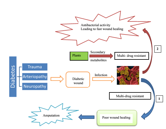
Diabetics are susceptible to severe trophic problems and wounds, which are frequently referred to as “unhealable wounds”. Untreated diabetic wounds provide an ideal habitat for the growth of bacteria, which can eventually develop resistance to standard treatments and need amputation. This study aimed to assess the in vitro antibacterial activity of ethanolic and aqueous extracts of Eriosema robustum leaves on the same bacterium, as well as the resistance profile of bacteria isolated from diabetic wounds to conventional antibiotics. Thirty samples from diabetic wounds were collected at the Baleveng Hospital, subjected to bacteria isolation and antibiotics resistance tests using the disk diffusion method. On the other hands, leaves of Eriosema robustum were collected, hydroethanolic extraction were performed. The extract obtained was used for the antibacterial (by determination of the MIC) and antioxidant (DPPH and FRAP tests) activities as well as phytochemicals composition (using HPLC methods) assessment. Amon the 30 samples analyzed, 19 (63.33 %) showed bacterial infections and Staphylococcus aureus was the most common pathogen (48 %), followed by Pseudomonas aeruginosa (24 %), Klebsiella pneumoniae (19 %), and Streptococcus agalactiae (9 %). Fifty percent (50 %) of S. aureus isolates were MRSA (methicillin-resistant S. aureus). The MIC value obtained 64 μg/mL against S. aureus, Streptococcus agalactiae and Pseudomonas aeruginosa. Phytochemicals analysis of fractions from the 70 ° hydroethanolic leaves of Eriosema robustum revealed the presence of coumarin, gallic acid, quinoline, vanillin, ascorbic acid, caffeic acid, kaempferol, and cinnamic acid. The antibacterial and antioxidant potential of the extracts highlighted in this study could make this plant a good avenue for the discovery of new molecules effective against infected diabetic wounds.
HIGHLIGHTS
- In this study, a total of 30 diabetic wounds were examined, and it was found that 63.33 % of them had infections. Staphylococcus aureus was the most frequently occurring bacteria, followed by Pseudomonas aeruginosa, Klebsiella pneumoniae, and Streptococcus agalactiae.
- The current study has revealed that, 96 % of the Staphylococcus aureus strains isolated were resistant to both tetracycline and methicillin, with the majority of the bacteria also showing resistance to amoxicillin.
- The hydroethanolic and aqueous extracts of Eriosema robustum displayed antibacterial activity against most of the isolated bacteria and the phytochemical analysis (HPLC) of fractions from the 70 ° hydroethanolic leaf extract revealed the presence of several secondary metabolites, including coumarin, gallic acid, quinoline, vanillin, ascorbic acid, caffeic acid, kaempferol, and cinnamic acid.
- This study opens up a new horizon in the management of diabetic wounds, and makes Eriosema robustum a potential candidate for the formulation of new drugs active against multi-resistant bacteria found in diabetic subjects.
GRAPHICAL ABSTRACT

Downloads
References
P Zhang, J Lu, Y Jing, S Tang, D Zhu and Y Bi. Global epidemiology of diabetic foot ulceration: A systematic review and meta-analysis. Ann. Med. 2017; 49, 106-16.
DG Armstrong, AJM Boulton and SA Bus. Diabetic foot ulcers and their recurrence. New Engl. J. Med. 2017; 376, 2367-75.
M Lepäntalo, J Apelqvist, C Setacci, JB Ricco, GD Donato, F Becker, H Robert-Ebadi, P Cao, HH Eckstein, PD Rango, N Diehm, J Schmidli, M Teraa, FL Moll, F Dick and AH Davies. Chapter V: Diabetic foot. Eur. J. Vasc. Endovasc. Surg. 2011; 42, 60-74.
E Chantelau, T Tanudjaja, F Altenhofer, Z Ersanli, S Lacigova and C Metzger. Antibiotic treatment for uncomplicated neuropathic forefoot ulcers in diabetes: A controlled trial. Diabet. Med. 1996; 13, 156-159.
BA Lipsky, É Senneville, ZG Abbas, J Aragón-Sánchez, M Diggle, JM Embil, S Kono, LA Lavery, M Malone, SAV Asten, V Urbančič-Rovan, EJG Peters and International Working Group on the Diabetic Foot. Guidelines on the diagnosis and treatment of foot infection in persons with diabetes (IWGDF 2019 update). Diabetes Metabol. Res. Rev. 2020; 36, e3280.
A Wright, S Wood, JD Silva and JS Bell. Systemic antimicrobial therapy for diabetic foot infections: An overview of systematic reviews. Antibiotics 2023; 12, 1041.
L Aceituno-Mata, J Tardío and M Pardo-de-Santayana. The persistence of flavor: Past and present use of wild food plants in Sierra Norte de Madrid, Spain. Front. Sustain. Food Syst. 2021; 4, 610238.
SH Zitouni-Nourine, N Belyagoubi-Benhammou, F El-Houaria Zitouni-Haouar, O Douahi, F Chenafi, H Fetati, SC Sari, A Benmahieddine, C Zaoui, FZN Mekaouche, FA Bekkara, N Kambouche, A Gismondi and H Toumi. Echinops spinosissimus turra root methanolic extract: Characterization of the bioactive components and relative wound healing, antimicrobial and antioxidant properties. Plants 2022; 11, 3440.
JB Gillett, RM Polhill and B Verdcourt. Flora of tropical East Africa. Leguminosae (part 4), subfamily papilionoideae (2), part 3. H Beentje, RM Polhill (Eds.). Crown Agents for Overseas Governments & Administrations,London, 1971.
JO Kokwaro. Medicinal plants of East Africa. University of Nairobi Press, Nairobi, Kenya, 2009.
MD Awouafack, LJ McGaw, S Gottfried, R Mbouangouere, P Tane and M Spiteller. Antimicrobial activity and cytotoxicity of the ethanol extract, fractions and eight compounds isolated from Eriosema robustum (Fabaceae). BMC Compl. Med. Ther. 2013; 13, 289.
SB Ateba, D Njamen and L Krenn. The genus eriosema (Fabaceae): From the ethnopharmacology to an evidence-based phytotherapeutic perspective? Front. Pharmacol. 2021; 12, 641225.
Clinical and Laboratory Standards Institute. Performance standards for antimicrobial susceptibility testing. Clinical and Laboratory Standards Institute, Pennsylvania, 2020.
GN Zirihi, AM Kra and F Guédé-Guina. Evaluation of the antifungal activity of microglossa pyrifolia (Larmarck) O. kuntze (Asteraceae) “Pymi” on the in vitro growth of candida albicans. Revue de Médecine et de Pharmacie 2003; 17, 11-9.
SPN Mativandlela, N Lall and JJM Meyer. Antibacterial, antifungal and antitubercular activity of Pelargonium reniforme (CURT) and Pelargonium sidoides (DC) (Geraniaceae) root extracts. S. Afr. J. Bot. 2006; 72, 232-7.
Mensor, FS Menezes, GG Leitao, ASO Reis, TCD Santos, CS Coube and SG Leitao. Screening of Brazilian plant extracts for antioxidant activity by the used DPPH free radical method. Phytother. Res. 2001; 15, 127-30.
IF Benzie and J Strain. The ferric reducing ability of plasma (FRAP) as a measure of “antioxidant power”: The FRAP assay. Anal. Biochem. 1996; 239, 70-6.
A Ramde-Tiendrebeogo, A Tibiri, A Hilou, M Lompo, H Millogo-Kone, OG Nacoulma and IP Guissou. Antioxidative and antibacterial activities of phenolic compounds from Ficus sue Forssk. Int. J. Biol. Chem. Sci. 2012; 6, 328-36.
C Chang, M Yang, H Wen and J Chern. Estimation of total flavonoid content in propolis by two complementary colorimetric methods. J. Food Drug Anal. 2002; 10, 178-82.
M Govindappa, SS Naga, MN Poojashri, TS Sadananda, CP Chandrappa, G Santoyo, P Sharanappa and NVA Kumar. Antimicrobial, antioxidant and in virto anti-inflammatory activity of ethanol extract and active phytochemical screening of Wedelia trilobata (L.). Hitchc. J. Med. Plants Res. 2011; 3, 43-51.
E Khadraoui, O Fendi, I Gaigi, N Trabelsi, A Trimech and MF Ben. Facteurs favorisants les lésions des pieds chez le diabétique. Diabetes Metabol. 2012; 32, A115.
KE Agbemadon. Bactériologie et antibiothérapie des plaies diabétiques dans le service de médecine interne d’hôpital du point G. Université des Sciences, des Techniques et des Technologies de Bamako, Bamako, Mali, 2015.
DP Abrogoua, A Bamba, E Doffou and A Lokrou. Évaluation économique de la prise en charge médicamenteuse du pied diabétique au CHU de Yopougon, Abidjan, Côte d’IvoireEconomic assessment of diabetic foot’s medication management in the teaching hospital of Yopougon, Abidjan, Ivory Coast. Médecine des Maladies Métaboliques 2019; 13, 91-5.
F Mauvais-Jarvis, NB Merz, PJ Barnes, RD Brinton, C Juan-Jesus, DL DeMeo, GJD Vries, CN Epperson, R Govindan, SL Klein, A Lonardo, PM Maki, LD McCullough, V Regitz-Zagrosek, JG Regensteiner, JB Rubin, K Sandberg and A Suzuki. Sex and gender: Modifiers of health, disease, and medicine. Lancet 2020; 396, 565-82.
A-S Vanherwegen, P Lauwers, A Lavens, K Doggen and E Dirinck.Sex differences in diabetic foot ulcer severity and outcome in Belgium. PLos One 2023; 18, e0281886.
A White, BD Sousa, RD Visser, R Hogston, SA Madsen and P Makara. The state of men’s health in Europe: Extended report. Directorate-General for Health and Consumers, European Union, 2011.
MA Rossaneis, MCFL Haddad, TAF Mathias and SS Marcon. Differences in foot self-care and lifestyle between men and women with diabetes mellitus. Revista Latino Americana De Enfermagem 2016; 24, e2761.
A Ho¨hn, J Gampe, R Lindahl-Jacobsen, K Christensen and A Oksuyzan. Do men avoid seeking medical advice? A register-based analysis of gender-specific changes in primary healthcare use after first hospitalisation at ages 60+ in Denmark. J. Epidemiol. Community Health 2020; 74, 573-9.
A Raliyatu, DG Ibrahim, EU Andrew and AR Mansur. Prevalence and risk factors of diabetes foot ulcers in Kano, northwestern Nigeria. Clin. Diabetes Endocrinol. 2023; 9, 6.
N Janice. Farmers with Diabetes: Risks fo lower extremity injury and disability. J Agromedicine 2000; 6, 59-68.
D Fomba. Etude épidémio-clinique des facteurs déclenchants la plaie du pied diabétique au service de médecine et d’endocrinologie de l’Hôpital du mali. Université des Sciences, des Techniques et des Technologies de Bamako, Bamako, Mali, 2022.
AL Oates, JMB Frank, B Andrew and AJ McBain. Molecular and culture-based assessment of the microbial diversity of diabetic chronic foot wounds and contralateral skin sites. J. Clin. Microbiol. 2012; 50, 2263-71.
SAS Hitam, SA Hassan and N Maning. The significant association between polymicrobial diabetic foot infection and its severity and outcomes. Malaysian J. Med. Sciences. 2019; 26, 107-14.
Yakout and Abdelwahab. Diabetic foot ulcer infectins and Pseudomonas aeruginosa biofim production during the covid-19 pandemic. J. Pure Appl. Microbiol. 2022; 16, 138-46.
S Farshad, R Ranjbar, M Anvarinejad, MA Shahidi and M Hosseini. Emergence of multi drug resistant strains of Eschetichia coli isolated from urinary tract infection. Open Conf. Proc. J. 2010; 1, 192-6.
U Trivedi, S Parameswaran, A Armstrong, D Burgueno-Vega, J Griswold, S Dissanaike and KP Rumbaugh. Prevalence of multiple antibiotic resistant infections in diabetic versus non diabetic wound. J. Pathogens 2014; 2014, 173053.
VC Bashige, AS Bakari, PN Okusa, EM Kalonda and JBS lumbu. Criblage phytochimique et activité antimicrobienne de six rhizomes comestibles utilisés en médecine traditionnelle à Lubumbashi (RDC). Int. J. Biol. Chem. Sci. 2020; 14, 1367-80.
D Gatsing and GI Adoga. AntiSalmonellal activity and screening of the various parts of Cassia petersiana bolle (caesalpiniaceae). Res. J. Microbiol. 2007; 2, 876-80.
JDD Tamokou, TA Mbaveng and V Kuete. Chapter 8 - Antimicrobial activities of African medicinal spices and vegetables. Med. Spices Vegetables Af. 2017; 2017, 207-37.
SK Prasad, D Laloo, M Kumar and S Hemalatha. Antidiarrhoeal evaluation of root extract, its bioactive fraction, and lupinifolin isolated from Eriosema chinense. Planta Med. 2013; 79, 1620-7.
MC Fomogne-Fodjo, VS Van, DT Ndinteh, RW Krause and DK Olivier. Antibacterial activities of plants from Central Africa used traditionally by the Bakola pygmies for treating respiratory and tuberculosis-related symptoms. J. Ethnopharmacol. 2014; 155, 123-31.
JM Gutteridge and B Halliwell. Free radicals and antioxidants in the year 2000. A historical look to the future. Ann. New York Acad. Sci. 2000; 899, 136-47.
A Arora, RK Sairam and GC Srivastave. Oxidative stress and anti-oxidative systems in plants. Curr. Sci. 2002; 82, 1227-38.
KRC Vijaya and DRM Sreeramulu. Antioxidant activity of fresh and dry fruits commonly consumed in India. Food Res. Int. 2010; 43, 285-8.
SE Drewes, MM Horn, F Khan, OQ Munro, JTB Dhlamini, C Rakuambo and JJM Meyer. Minor pyrano-isoflavones from Eriosema kraussianum: Activity-, structure-, and chemical reaction studies. Phytochemistry 2004; 65, 1955-61.
MD Awouafack, SF Kouam, H Hussain, D Ngamga, P Tane, B Schulz, IR Green and K Krohn. Antimicrobial prenylated dihydrochalcones from Eriosema glomerata. Planta Med. 2008; 74, 50-4.
S Sutthivaiyakit, O Thongnak, T Lhinhatrakool, O Yodchun, R Srimark, P Dowtaisong and M Chuankamnerdkarn. Cytotoxic and antimycobacterial prenylated flavonoids from the roots of Eriosema chinense. J. Nat. Prod. 2009; 72, 1092-6.
D Savoia. Plant derived antimicrobial compounds. Future Microbiol. 2012; 7, 979¬990.
Downloads
Published
How to Cite
Issue
Section
License
Copyright (c) 2024 Walailak University

This work is licensed under a Creative Commons Attribution-NonCommercial-NoDerivatives 4.0 International License.


