
The Relationship between the Change in Pelvic Alignment during Moderate-Intensity Running and the Subjective Rating of Perceived Exertion, Rating of Perceived Exertion for Legs and Gluteus Muscles Fatigue in Female Novice and Recreational Runners
DOI:
https://doi.org/10.48048/tis.2024.7286Keywords:
Pelvic drop, Pelvic alignment, Moderate-intensity runningAbstract
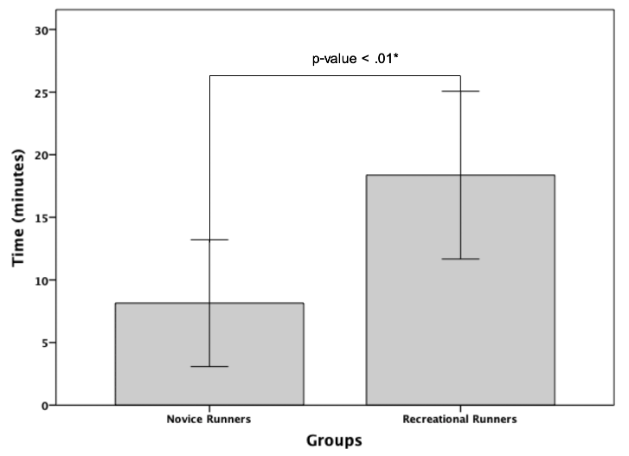
Running has become 1 of the 2 most well-known exercises for health; however, running may cause injuries. The purpose of this analytical descriptive study was to examine the occurrence of pelvic drop during moderate-intensity running in female novice and recreational runners; as well as investigating the relationship between the change in pelvic alignment and Rating of Perceived Exertion (RPE), Rating of Perceived Exertion for Legs (RPElegs) and Gluteus muscles fatigue in novice and recreational runners. The participants, aged between 18 and 35 years old, comprised 27 novice runners (less than 1 year of running experience) and 27 recreational runners (2 - 4 years running experience). The participants in both groups performed the maximal oxygen uptake (VO2max) test and ran at a self-selected speed on a treadmill for 30 min at moderate-intensity within the specified heart rate range of 40 - 59 % heart rate reserve (HRR). RPE, RPElegs and sEMG were collected every 2 min and the pelvic kinematic data were also recorded every minute during the 30 min treadmill running test. The data were analyzed with a statistical significance level at p-value < 0.05. The results demonstrated that the time in starting pelvic drop was 8.15 ± 5.07 and 18.37 ± 6.70 min in female novice and recreational runner groups, respectively (p < 0.01). Pearson’s correlations showed relationship between the pelvic alignment and RPE in novice runners (r = 0.47, p < 0.01) and recreational runners (r = 0.39, p < 0.01). Besides, Pearson’s correlations showed relationship between the pelvic alignment and RPElegs in novice runners (r = 0.48, p < 0.01) and recreational runners (r = 0.32, p < 0.01). In both groups, there was no relationship between the pelvic alignment and Gluteus medius and Gluteus maximus muscle fatigue. According to the study, the occurrence of a pelvic drop in novice runners was faster than recreational runners. The study indicated that consideration of RPE and RPElegs variables might be beneficial for female runners and future studies.
HIGHLIGHTS
- The occurrence of pelvic drop in novice runners was faster than recreational runners.
- There was a relationship between the change in pelvic alignment and RPE and also RPElegs in female novice and recreational runners.
- The study indicated that consideration of RPE and RPElegs variables might be beneficial for female runners and future studies.
GRAPHICAL ABSTRACT

Downloads
References
KB Watson, GM Frederick, CD Harris, SA Carlson and JE Fulton. US adults’ participation in specific activities: Behavioral risk factor surveillance system - 2011. J. Phys. Activ. Health 2015; 12, S3-S10.
WL Haskell, IM Lee, RR Pate, KE Powell, SN Blair, BA Franklin, CA Macera, GW Heath, PD Thompson and A Bauman. Physical activity and public health: Updated recommendation for adults from the American College of Sports Medicine and the American Heart Association. Circulation 2007; 116, 1081.
JD Cantwell. Cardiovascular aspects of running. Clin. Sports Med. 1985; 4, 627-40.
American College of Sports Medicine. ACSM’s guidelines for exercise testing and prescription. by LWW, Pennsylvania, 2017.
RO Nielsen, I Buist, H Sørensen, M Lind and S Rasmussen. Training errors and running related injuries: A systematic review. Int. J. Sports Phys. Ther. 2012; 7, 58-75.
BE Smith, J Selfe, D Thacker, P Hendrick, M Bateman, F Moffatt, MS Rathleff, TO Smith and P Logan. Incidence and prevalence of patellofemoral pain: A systematic review and meta-analysis. PloS One 2018; 13, e0190892.
L Malisoux, N Chambon, N Delattre, N Gueguen, A Urhausen and D Theisen. Injury risk in runners using standard or motion control shoes: A randomised controlled trial with participant and assessor blinding. Br. J. Sports Med. 2016; 50, 481-7.
D Theisen, L Malisoux, J Genin, N Delattre, R Seil and A Urhausen. Influence of midsole hardness of standard cushioned shoes on running-related injury risk. Br. J. Sports Med. 2014; 48, 371-6.
PE Niemuth, RJ Johnson, MJ Myers and TJ Thieman. Hip muscle weakness and overuse injuries in recreational runners. Clin. J. Sport Med. 2005; 15, 14-21.
MB Pohl, DR Mullineaux, CE Milner, J Hamill and IS Davis. Biomechanical predictors of retrospective tibial stress fractures in runners. J. Biomech. 2008; 41, 1160-5.
J Becker, S James, R Wayner, L Osternig and LS Chou. Biomechanical factors associated with Achilles tendinopathy and medial tibial stress syndrome in runners. Am. J. Sports Med. 2017; 45, 2614-21.
JJ Hannigan. 2014, The relationship between hip strength and hip, pelvis, and trunk kinematics in healthy runners. Ph. D. Dissertation. University of Oregon, Oregon, United States.
ML Ireland, JD Willson, BT Ballantyne and IM Davis. Hip strength in females with and without patellofemoral pain. J. Orthop. Sports Phys. Ther. 2003; 33, 671-6.
MR Prins and PVD Wurff. Females with patellofemoral pain syndrome have weak hip muscles: A systematic review. Aust. J. Physiother. 2009; 55, 9-15.
R Ferber, B Noehren, J Hamill and I Davis. Competitive female runners with a history of iliotibial band syndrome demonstrate atypical hip and knee kinematics. J. Orthop. Sports Phys. Ther. 2010; 40, 52-8.
C Dunphy, S Casey, A Lomond and D Rutherford. Contralateral pelvic drop during gait increases knee adduction moments of asymptomatic individuals. Hum. Mov. Sci. 2016; 49, 27-35.
MG Gazendam and AL Hof. Averaged EMG profiles in jogging and running at different speeds. Gait Posture 2007; 25, 604-14.
RW Willy, KT Manal, EE Witvrouw and IS Davis. Are mechanics different between male and female runners with patellofemoral pain? Med. Sci. Sports Exerc. 2012; 44, 2165-71.
C Bramah, SJ Preece, N Gill and L Herrington. Is there a pathological gait associated with common soft tissue running injuries? Am. J. Sports Med. 2018; 46, 3023-31.
EN Burnet. 2008, Frontal plane pelvic drop in runners: Causes and clinical implications. Ph. D. Dissertation. Virginia Commonwealth University, Virginia, United States.
L Linton and S Valentin. Running with injury: A study of UK novice and recreational runners and factors associated with running related injury. J. Sci. Med. Sport 2018; 21, 1221-5.
D Gerasimuk, E Malchrowicz-Mośko, A Stanula, E Bezuglov, E Achkasov, A Swinarew and Z Waśkiewicz. Age-related differences in motivation of recreational runners, marathoners, and ultra-marathoners. Front. Psychol. 2021; 12, 738807.
S Sabharwal and A Kumar. Methods for assessing leg length discrepancy. Clin. Orthop. Relat. Res. 2008; 466, 2910-22.
G Borg. Borg’s perceived exertion and pain scales. Human Kinetics, Illinois, 1998.
M Garnacho-Castaño, R Domínguez and J Maté-Muñoz. Understanding the meaning of lactate threshold in resistance exercises. Int. J. Sports Med. 2015; 36, 371-7.
C Easton, S Turner and YP Pitsiladis. Creatine and glycerol hyperhydration in trained subjects before exercise in the heat. Int. J. Sport Nutr. Exerc. Metabol. 2007; 17, 70-91.
AM Brown, RA Zifchock, M Lenhoff, J Song and HJ Hillstrom. Hip muscle response to a fatiguing run in females with iliotibial band syndrome. Hum. Mov. Sci. 2019; 64, 181-90.
C Jacobs, TL Uhl, M Seeley, W Sterling and L Goodrich. J. Athletic Train. 2005; 40, 203.
EN Burnet and PE Pidcoe. Isometric gluteus medius muscle torque and frontal plane pelvic motion during running. J. Sports Sci. Med. 2009; 8, 284-8.
C Foster. VO2 max and training indices as determinants of competitive running performance. J. Sports Sci. 1983; 1, 13-22.
C Ritchie. Rating of perceived exertion (RPE). J. Physiother. 2012; 58, 62.
H Zhao, T Nishioka and J Okada. Validity of using perceived exertion to assess muscle fatigue during resistance exercises. PeerJ 2022; 10, e13019.
H Zhao, D Seo and J Okada. Validity of using perceived exertion to assess muscle fatigue during back squat exercise. BMC Sports Sci. Med. Rehabil. 2023; 15, 14.
AC Cruz, ST Fonseca, VL Araújo, DS Carvalho, LD Barsante, VA Pinto and TR Souza. Pelvic drop changes due to proximal muscle strengthening depend on foot-ankle varus alignment. Appl. Bionics Biomech. 2019; 2019, 2018059.
Downloads
Published
How to Cite
Issue
Section
License
Copyright (c) 2023 Walailak University

This work is licensed under a Creative Commons Attribution-NonCommercial-NoDerivatives 4.0 International License.


