
The Optimal Dose of the Green Tea and Coffee Extracts to Suppress the Expression of PPAR-γ and C/EBP-α on Differentiated 3T3-L1 Adipocytes
DOI:
https://doi.org/10.48048/tis.2024.7182Keywords:
C/EBP-α, Green tea, Green coffee, Obesity, PPAR-γ, 3T3-L1Abstract
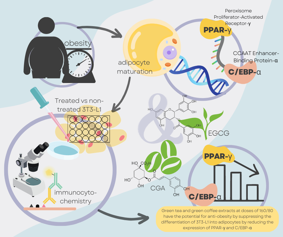
Obesity is the excess fat content in the body caused by the expansion of white adipose tissue. This condition begins with the differentiation of adipose tissue that is known to be controlled by 2 main transcription factors, such as Peroxisome Proliferator-Activated Receptor-γ (PPAR-γ) and CCAAT Enhancer-Binding Protein-α (C/EBP-α). Their activation and collaboration are critical for developing functioning adipocytes and preserving metabolic balance in adipose tissue. Research on the benefits of natural bioactive components that regulate adipogenesis has recently become an exciting focus. Foodstuffs reported to affect this condition positively include green coffee and green tea. The primary substance in green coffee is chlorogenic acid (CGA), meanwhile in green tea is epigallocatechin gallate (EGCG). Based on medical benefit potential, the present study evaluated the dose that produced the best effect on PPAR-γ and C/EBP-α expression between single and combined doses compared to undifferentiated adipocytes (3T3-L1). 3T3-L1 were cultured and divided into negative (NEG) and positive (DIF) groups. The DIF group is obtained by induction of differentiation, then divided into seventeen therapeutic doses. At the end of therapy, cells were fixed to measure the expression of PPAR-γ and C/EBP-α using the immunocytochemistry method. The DIF group produced the highest expression of PPAR-γ and C/EBP-α. Among the single-dose group, the lowest PPAR-γ was found in C320 and C/EBP-α in the T320 group. Meanwhile, the dose with the lowest PPAR-γ and C/EBP-α expression was found in the combination of green tea and coffee (TC) 160/80 (p-value = 0.00). The findings of this study showed that a combination of green tea and coffee extracts at doses of 160/80 has the potential for anti-obesity by suppressing the differentiation of 3T3-L1 into adipocytes by reducing the expression of PPAR-γ and C/EBP-α.
HIGHLIGHTS
- Obesity is the excess fat content in the body caused by the expansion of white adipose tissue. This condition begins with the differentiation of adipose tissue that is known to be controlled by Peroxisome Proliferator-Activated Receptor-γ (PPAR-γ) and CCAAT Enhancer-Binding Protein-α (C/EBP-α)
- Natural bioactive components that regulate adipogenesis in green coffee are chlorogenic acid (CGA) and epigallocatechin gallate found in green tea (EGCG). Based on this potential, the present study evaluated the dose that produced the best effect on PPARɣ and C/EBP-α protein expression between single and combined doses compared to undifferentiated adipocytes (3T3-L1)
- Based on this study, the differentiated group of adipocytes (DIF) produced the highest expression of PPAR-γ and C/EBP-α. Among the single dose group, which showed the lowest average value of PPARɣ was C320, and for C/EBP-α was the T320 group. Meanwhile, the dose with the lowest PPAR-γ and C/EBP-α expression came from the combination of green tea and coffee (TC) 160/80 (p-value = 0.00)
- So, this research shows that green tea and green coffee extract significantly affected the expression of PPAR-γ and C/EBP-α. Green tea and coffee extracts at doses of TC 160/80 have the potential for anti-obesity by suppressing the differentiation of 3T3-L1 into adipocytes by reducing the expression of PPAR-γ and C/EBP-α
GRAPHICAL ABSTRACT
Downloads
References
World Health Organization. Obesity and overweight, Available at https://www.who.int/news-room/fact-sheets/detail/obesity-and-overweight, accessed February 2023.
BM Popkin, LS Adair and SW Ng. Global nutrition transition and the pandemic of obesity in developing countries. Nutr. Rev. 2012; 70, 3-21.
JM Poti, B Braga and B Qin. Ultra-processed food intake and obesity: What really matters for health-processing or nutrient content? Curr. Obes. Rep. 2017; 6, 420-31.
S Camacho and A. Ruppel. Is the calorie concept a real solution to the obesity epidemic? Glob. Health Action 2017; 10, 1289650.
X Lin and H Li. Obesity: Epidemiology, pathophysiology, and therapeutics. Front. Endocrinol. 2021; 12, 706978.
JQ Purnell. Definitions, classification, and epidemiology of obesity, Available at https://www.ncbi.nlm.nih.gov/books/NBK279167, accessed, February 2023.
NHS. Obesity, Available at https://www.nhs.uk/conditions/obesity/, accessed February 2023.
FS Mohammed, A Ghosh, S Pal, C Das, SY Alomar, M Patwekar, F Patwekar, BH Jeon and F Islam. Hydroalcoholic Extract of Sechium edule fruits attenuates QT prolongation in high fat diet-induced hyperlipidemic mice. Evid. Based Complement. Alternat. Med. 2022; 2022, 8682316.
SM Grundy. Adipose tissue and metabolic syndrome: Too much, too little or neither. Eur. J. Clin. Invest. 2015; 45, 1209-17.
HN Ginsberg and PR MacCallum. The obesity, metabolic syndrome, and type 2 diabetes mellitus pandemic: Part I. Increased cardiovascular disease risk and the importance of atherogenic dyslipidemia in persons with the metabolic syndrome and type 2 diabetes mellitus. J. Cardiometab. Syndr. 2009; 4, 113-9.
DR Leitner, G Frühbeck, V Yumuk, K Schindler, D Micic, E Woodward and H Toplak. Obesity and type 2 diabetes: Two diseases with a need for combined treatment strategies - EASO can lead the way. Obes. Facts 2017; 10, 483-92.
DC Lau and H Teoh. Current and Emerging Pharmacotherapies for weight management in prediabetes and diabetes. Can. J. Diabetes 2015; 39, S134-S141.
A Quazi, FP Mohsina, IP Faheem and S Priya. In silico ADMET analysis, molecular docking and in vivo anti diabetic activity of polyherbal tea bag formulation in Streptozotocin-nicotinamide induced diabetic rats. Int. J. Health Sci. 2022; 6, 343-72.
S Sugii, P Olson, DD Sears, M Saberi, AR Atkins, GD Barish, SH Hong, GL Castro, YQ Yin, MC Nelson, G Hsiao, DR Greaves, M Downes, RT Yu, JM Olefsky and RM Evans. PPARɣ activation in adipocytes is sufficient for systemic insulin sensitization. Proc. Natl. Acad. Sci. Unit. States Am. 2009; 106, 22504-9.
M Guo, C Li, Y Lei, S Xu, D Zhao and XY Lu. Role of the adipose PPARγ-adiponectin axis in susceptibility to stress and depression/anxiety-related behaviors. Mol. Psychiatr. 2017; 22, 1056-68.
L Guo, X Li and QQ Tang. Transcriptional regulation of adipocyte differentiation: A central role for CCAAT/enhancer-binding protein (C/EBP) β. J. Biol. Chem. 2015; 290, 755-61.
HH Cho, SJ Lee, SH Kim, SH Jang, C Won, HD Kim, TH Kim and JH Cho. Acer tegmentosum Maxim inhibits adipogenesis in 3t3-l1 adipocytes and attenuates lipid accumulation in obese rats fed a high-fat diet. Nutrients 2020; 1, 3753.
M Longo, F Zatterale, J Naderi, L Parrillo, P Formisano, GA Raciti, F Beguinot and C Miele. Adipose tissue dysfunction as determinant of obesity-associated metabolic complications. Int. J. Mol. Sci. 2019; 20, 2358.
C Sun, S Mao, S Chen, W Zhang and C Liu. PPARs-orchestrated Metabolic homeostasis in the adipose tissue. Int. J. Mol. Sci. 2021; 22, 8974.
QA Wang, F Zhang, L Jiang, R Ye, Y An, M Shao, C Tao, RK Gupta and PE Scherer. Peroxisome proliferator-activated receptor γ and its role in adipocyte homeostasis and thiazolidinedione-mediated insulin sensitization. Mol. Cell. Biol. 2018; 38, e00677-17.
N Saraf, PK Sharma, SC Mondal, VK Garg and AK Singh. Role of PPARg2 transcription factor in thiazolidinedione-induced insulin sensitization. J. Pharm. Pharmacol. 2012; 64, 161-71.
ED Rosen, CH Hsu, X Wang, S Sakai, MW Freeman, FJ Gonzalez and BM Spiegelman. C/EBPalpha induces adipogenesis through PPARgamma: A unified pathway. Genes Dev. 2002; 16, 22-6.
MI Lefterova, Y Zhang, DJ Steger, M Schupp, J Schug, A Cristancho, D Feng, D Zhuo, CJ Stoeckert Jr, XS Liu and MA Lazar. PPARgamma and C/EBP factors orchestrate adipocyte biology via adjacent binding on a genome-wide scale. Genes Dev. 2008; 22, 2941-52.
TC Otto and MD Lane. Adipose development: from stem cell to adipocyte. Crit. Rev. Biochem. Mol. Biol. 2005; 40, 229-42.
N Yokomori, M Tawata and T Onaya DNA demethylation during the differentiation of 3T3-L1 cells affects the expression of the mouse GLUT4 gene. Diabetes 1999; 48, 685-90.
TA Pedersen, O Bereshchenko, S Garcia-Silva, O Ermakova, E Kurz, S Mandrup, BT Porse and C Nerlov. Distinct C/EBPalpha motifs regulate lipogenic and gluconeogenic gene expression in vivo. EMBO J. 2007; 26, 1081-93.
International Coffee Organization. Monthly coffee market report, Available at http://www.ico.org/documents/cmr-0712-e.pdf, accessed February 2023.
C Cabrera, R Artacho and R Gime. Beneficial effects of green tea - a review. J. Am. Coll. Nutr. 2006; 25, 79-99.
SM Chacko, PT Thambi, R Kuttan and I Nishigaki. Beneficial effects of green tea: A literature review. Chin. Med. 2010; 5, 13.
A Rojas-González, CY Figueroa-Hernández, O González-Rios, ML Suárez-Quiroz, RM González-Amaro, ZJ Hernández-Estrada and P Rayas-Duarte. Coffee chlorogenic acids incorporation for bioactivity enhancement of foods: A review. Molecules 2022; 27, 3400.
SE Park, JH Choi, HJ Lee, K Seo and S Kim. Anti-adipogenic effect of chlorogenic acid in 3T3-L1 Adipocytes. In: Proceedings of the Plant Resources Society of Korea Conference, South Korea. 2018.
M Sano, M Tabata, M Suzuki, M Degawa, T Miyase and M Maeda-Yamamoto. Simultaneous determination of twelve tea catechins by high-performance liquid chromatography with electrochemical detection. Analyst 2001; 126, 816-20.
JB Seo, HM Moon, WS Kim, YS Lee, HW Jeong, EJ Yoo, J Ham, H Kang, MG Park, KR Steffensen, TM Stulnig, JA Gustafsson, SD Park and JB Kim. Activated liver X receptors stimulate adipocyte differentiation through induction of peroxisome proliferator-activated receptor gamma expression. Mol. Cell. Biol. 2004; 24, 3430-44.
H Kim and K Sakamoto. (-)-Epigallocatechin gallate suppresses adipocyte differentiation through the MEK/ERK and PI3K/Akt pathways. Cell Biol. Int. 2012; 36, 147-53.
PF Hung, BT Wu, HC Chen, YH Chen, CL Chen, MH Wu, HC Liu, MJ Lee and YH Kao. Antimitogenic effect of green tea (-)-epigallocatechin gallate on 3T3-L1 preadipocytes depends on the ERK and Cdk2 pathways. Am. J. Physiol. Cell Physiol. 2005; 288, C1094-C1108.
AG Fischer and PM Kummer. 1993, Process for decaffeinating raw coffee, U.S. Patent 5208056A.
H Liang, Y Liang, J Dong J, Lu, H Xu and H Wang. Decaffeination of fresh green tea leaf (Camellia Sinensis) by hot water treatment. Food Chem. 2002; 101, 1451-6.
M Lukitasari, D Nugroho, M Rohman, N Widodo, A Farmawati and P Hastuti. Beneficial effects of green coffee and green tea extract combination on metabolic syndrome improvement by affecting ampk and ppar-α gene expression. J. Advan. Pharmaceut. Tech. Res. 2020; 11, 81-5.
M Lukitasari, MS Rohman, DA Nugroho, NA Wahyuni, MN Kholis and N Widodo. Improvement of cardiac fibrosis biomarkers through inflammation inhibition by green tea and decaffeinated light roasted green coffee extract combination administration in metabolic syndrome rat model. F1000Research 2021; 10, 1013.
MS Rohman, M Lukitasari, DA Nugroho, R Ramadhiani, N Widodo, I Kusumastuty, NIP Nugrahini. Decaffeinated light-roasted green coffee and green tea extract combination improved metabolic parameters and modulated inflammatory genes in metabolic syndrome rats. F1000Research 2021; 10, 467.
AE Horvai, JT Schaefer, EK Nakakura and RJ O’Donnell. Immunostaining for peroxisome proliferator gamma distinguishes dedifferentiated liposarcoma from other retroperitoneal sarcomas. Mod. Pathol. 2008; 21, 517-24.
SR Farmer. Transcriptional control of adipocyte formation. Cell. Metab. 2006; 4, 263-73.
E Rosen, J Eguchi and Z Xu. Transcriptional targets in adipocyte biology. Expert Opin. Ther. Targets 2009; 13, 975-86.
ZH Li, R Carraro, RI Gregerman and DC Lau. Adipocyte differentiation factor (ADF): A protein secreted by mature fat cells that induces preadipocyte differentiation in culture. Cell Biol. Int. 1998; 22, 253-70.
DP Ramji and P Foka. CCAAT/enhancer-binding proteins: Structure, function and regulation. Biochem J. 2002; 365, 561-75.
GJ Darlington, SE Ross and OA MacDougald. The role of C/EBP genes in adipocyte differentiation. J. Biol. Chem. 1998; 273, 30057-60.
X Wang and C Hai. Redox modulation of adipocyte differentiation: hypothesis of “Redox Chain” and novel insights into intervention of adipogenesis and obesity. Free Radic. Biol. Med. 2015; 89, 99-125.
YK Park, L Wang, A Giampietro, B Lai, JE Lee and K Ge. Distinct Roles of Transcription Factors KLF4, Krox20, and Peroxisome Proliferator-Activated Receptor γ in adipogenesis. Mol. Cell Biol. 2017; 37, e00554-16.
HG Linhart, K Ishimura-Oka, F Demayo, T Kibe, D Repka, B Poindexter and GJ Darlington. C/EBPα is required for differentiation of white but not brown adipose tissue. Proc. Natl. Acad. Sci. Unit. States Am. 2001; 98, 12532-7.
SO Freytag, DL Paielli and JD Gilbert. Ectopic expression of the CCAAT/enhancer-binding protein alpha promotes the adipogenic program in a variety of mouse fibroblastic cells. Genes Dev. 1994; 8, 1654-63.
MS Madsen, R Siersbæk, M Boergesen, R Nielsen, and S Mandrup. Peroxisome proliferator-activated receptor γ and C/EBPα synergistically activate key metabolic adipocyte genes by assisted loading. Mol. Cell Biol. 2014; 34, 939-54.
T Furuyashiki, H Nagayasu, Y Aoki, H Bessho, T Hashimoto, K Kanazawa and H Ashida. Tea catechin suppresses adipocyte differentiation accompanied by down-regulation of PPARgamma2 and C/EBPalpha in 3T3-L1 cells. Biosci. Biotechnol. Biochem. 2004; 68, 2353-9.
FM Gregorie, CM Smas and HS Sul. Understanding adipocyte differentiation. Physiol. Rev. 1998; 78, 783-809.
MS Madsen, R Siersbæk, M Boergesen, R Nielsen and S Mandrup. Peroxisome proliferator-activated receptor γ and C/EBPα synergistically activate key metabolic adipocyte genes by assisted loading. Mol. Cell Biol. 2014; 34, 939-54.
X Yang, L Yin, T Li and Z Chen. Green tea extracts reduce adipogenesis by decreasing expression of transcription factors C/EBPα and PPARγ. Int. J. Clin. Exp. Med. 2014; 7, 4906-14.
W Lao, Y Tan, X Jin, L Xiao, JJ Kim and X Qu. Comparison of cytotoxicity and the anti-adipogenic effect of green tea polyphenols with Epigallocatechin-3-Gallate in 3T3-L1 Preadipocytes. Am. J. Chin. Med. 2015; 43, 1177-90.
K Sirichaiwetchakoon, GM Lowe, K Thumanu and G Eumkeb. The effect of Pluchea indica (L.) less. Tea on adipogenesis in 3T3-L1 adipocytes and lipase activity. Evid. Based Complement Alternat. Med. 2018; 2018, 4108787.
CC Kao, BT Wu, YW Tsuei, LJ Shih, YL Kuo and YH Kao. Green tea catechins: Inhibitors of glycerol-3-phosphate dehydrogenase. Planta Med. 2010; 76, 694-6.
C Maki, M Funakoshi-Tago, R Aoyagi, F Ueda, M Kimura, K Kobata, K Tago and H Tamura. Coffee extract inhibits adipogenesis in 3T3-L1 preadipocyes by interrupting insulin signaling through the downregulation of IRS1. PLoS One 2017; 12, e0173264.
M Carlström and SC Larsson. Coffee consumption and reduced risk of developing type 2 diabetes: A systematic review with meta-analysis. Nutr. Rev. 2018; 76, 395-417.
A Muley, P Muley and M Shah. Coffee to reduce risk of type 2 diabetes? A systematic review. Curr. Diabetes Rev. 2012; 8, 162-8.
R Aoyagi, M Funakoshi-Tago, Y Fujiwara and H Tamura. Coffee inhibits adipocyte differentiation via inactivation of PPARγ. Biol. Pharm. Bull. 2014; 37, 1820-5.
R Farias-Pereira, CS Park and Y Park. Mechanisms of action of coffee bioactive components on lipid metabolism. Food Sci. Biotechnol. 2019; 28, 1287-96.
SG Peng, YL Pang, Q Zhu, JH Kang, MX Liu and Z Wang. Chlorogenic acid functions as a novel agonist of PPARγ2 during the differentiation of mouse 3T3-L1 preadipocytes. Biomed Res. Int. 2018; 2018, 8594767.
WY Gao, PY Chen, HJ Hsu, CY Lin, MJ Wu and JH Yen. Tanshinone IIA downregulates lipogenic gene expression and attenuates lipid accumulation through the modulation of LXRα/SREBP1 Pathway in HepG2 Cells. Biomedicines 2021; 9, 326.
M Rebollo-Hernanz, Q Zhang, Y Aguilera, MA Martín-Cabrejas and EGD Mejia. Relationship of the phytochemicals from coffee and cocoa by-products with their potential to modulate biomarkers of metabolic syndrome in vitro. Antioxidants 2019; 8, 279.
J Taïlé, M Bringart, C Planesse, J Patché, P Rondeau, B Veeren, P Clerc, A Gauvin-Bialecki, S Bourane, O Meilhac, D Couret and MP Gonthier. Antioxidant polyphenols of antirhea borbonica medicinal plant and caffeic acid reduce cerebrovascular, inflammatory and metabolic disorders aggravated by high-fat diet-induced obesity in a mouse model of stroke. Antioxidants 2022; 11, 858.
Downloads
Published
How to Cite
Issue
Section
License
Copyright (c) 2023 Walailak University

This work is licensed under a Creative Commons Attribution-NonCommercial-NoDerivatives 4.0 International License.


