
Enhanced Osteogenic Differentiation of Human Mesenchymal Stem Cells with Synthetic Coral Matrix in the Presence or Absence of Growth Factors from the Platelet Rich-rich Plasma
DOI:
https://doi.org/10.48048/tis.2024.7168Keywords:
Synthetic coral matrix (SCM), Scaffold, Growth factors, Platelet rich plasma (PRP), Human mesenchymal stem cells (h-MSCs), Bone tissue engineeringAbstract
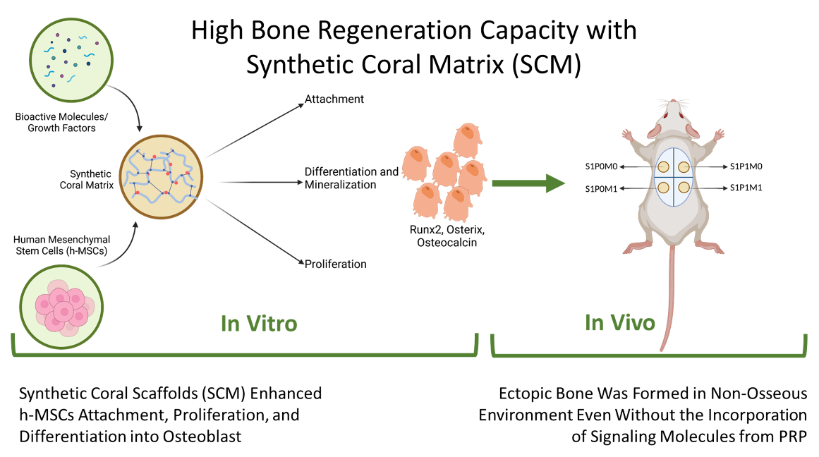
To solve problems in using natural sea corals with high biocompatibility, good osteoconductivity, and ideal degradability, a synthetic three-dimensional coral matrix was fabricated and observed for its capability to enhance osteogenic differentiation of human mesenchymal stem cells (h-MSCs), in the presence or absence of growth factors (GFs) supplied from platelet-rich plasma (PRP). Expressions of runx, osterix, and osteocalcin were investigated, following cell attachment, and proliferation analysis. Ectopic bone formation in nonosseous tissues of Sprague-Dawley rats at predetermined time intervals was investigated, including mineralized tissue growth in vivo. The 3D synthetic coral matrix (SCM) can interact with the GF cocktail in the PRP and MSCs to generate and secret bone extracellular matrix (ECM) both in vitro and in vivo. The matrix supplied with GF cocktail from the PRP provided an ideal microenvironment for MSCs to attach, proliferate, and differentiate into osteoblast faster, as indicated by the expression levels of runx2, osterix, and osteocalcin. The high capability of SCMs to enhance bone formation has been proven by the formation of ectopic bones in the nonosseous environment. The incorporated PRP provided blood proteins such as fibrin to slow down matrix degradation, whereas GF supplied by the PRP stimulated h-MSCs to attach and proliferate onto the matrix. Moreover, the GF supplied by the PRP enhanced osteogenic differentiation and mineralization, accelerating bone regeneration. Valorization phases are needed to apply the SCM for bone tissue engineering in clinics.
HIGHLIGHTS
- A synthetic coral matrix (SCM) with ideal macro- and microstructural appearance and physical and chemical properties, with tunable platelet-rich plasma (PRP)-loading capacity and release manner, was successfully fabricated
- The matrix provided an ideal microenvironment for human mesenchymal stem cells (h-MSCs) to attach, proliferate, and differentiate into osteoblasts indicated by high expression levels of bone regeneration markers
- The high capability of SCM to enhance bone regeneration has been validated by the formation of ectopic bones in the nonosseous environment of subcutaneous rat dorsal tissues
- The incorporation of a growth factor cocktail from the PRP did not alter the results in the matrix without growth factors, which act as signaling molecules
- The SCM was found to degrade faster in the group without h-MSCs, indicating that the system with cells promoted faster bone formation
GRAPHICAL ABSTRACT
Downloads
References
Y Tabata. Biomaterial technology for tissue engineering applications. J. R. Soc. Interface 2009; 6, S311-S324.
BP Chan and KW Leong. Scaffolding in tissue engineering: General approaches and tissue-specific considerations. Eur. Spine J. 2008; 17, 467-79.
SK Nandi, B Kundu, J Mukherjee, A Mahato, S Datta and VK Balla. Converted marine coral hydroxyapatite implants with growth factors: In vivo bone regeneration. Mater. Sci. Eng. C 2015; 49, 816-23.
D Srivastava and ND Witt. In vivo cellular reprogramming: The next generation. Cell 2016; 166, 1386-96.
CM Andreasen, JM Delaisse, BCVD Eerden, JPV Leeuwen, M Ding and TL Andersen. Understanding age-induced cortical porosity in women: The accumulation and coalescence of eroded cavities upon existing intracortical canals is the main contributor. J. Bone Miner. Res. 2018; 33, 606-20.
AI Birkhold, H Razi, R Weinkamer, GN Duda, S Checa and BM Willie. Monitoring in vivo (re) modeling: A computational approach using 4D micro-CT data to quantify bone surface movements. Bone 2015; 75, 210-21.
P Chocholata, V Kulda and Babuska. Fabrication of scaffolds for bone-tissue regeneration. Materials 2019; 12, 568.
H Pereira, IF Cengiz, FR Maia, F Bartolomeu, JM Oliveira, RL Reis and FS Silva. Physicochemical properties and cytocompatibility assessment of non-degradable scaffolds for bone tissue engineering applications. J. Mech. Behav. Biomed. Mater. 2020; 112, 103997.
ID Ana. Bone substituting materials in dental implantology. In: AS Budihardja and T Mücke (Eds.). Bone management in dental implantology. Springer, Cham, Switzerland, 2019, p. 121-4.
L Guo, Z Liang, L Yang, W Du, T Yu, H Tang, C Li and H Qiu. The role of natural polymers in bone tissue engineering. J. Contr. Release 2021; 338, 571-82.
N Aslankoohi, D Mondal, AS Rizkalla and K Mequanint. Bone repair and regenerative biomaterials: Towards recapitulating the microenvironment. Polymers 2019; 11, 1437.
R Shi, Y Huang, C Ma, C Wu and W Tian. Current advances for bone regeneration based on tissue engineering strategies. Front Med. 2019; 13, 160-88.
P Wang, L Zhao, J Liu, MD Weir, X Zhou and HH Xu. Bone tissue engineering via nanostructured calcium phosphate biomaterials and stem cells. Bone Res. 2014; 30, 14017.
Y Qian, Q Han, W Chen, J Song, X Zhao, Y Ouyang, W Yuan and C Fan. Platelet-rich plasma derived growth factors contribute to stem cell differentiation in musculoskeletal regeneration. Front Chem. 2017; 5, 89.
ID Ana. Hybrid biomaterials in drug delivery and biomedical applications. In: S Jana and S Jana (Eds.). Functional Biomaterials. Springer Nature, Singapore, 2022, p. 409-34.
R Hou, F Chen, Y Yang, X Cheng, Z Gao, HO Yang, W Wu and T Mao. Comparative study between coral-mesenchymal stem cells-rhBMP-2 composite and auto-bone-graft in rabbit critical-sized cranial defect model. J. Biomed. Mater. Res. A 2007; 80, 85-93.
AS Neto and JMF Ferreira. Synthetic and marine-derived porous scaffolds for bone tissue engineering. Materials 2018; 11, 1702.
KA Al-Salihi. In vitro evaluation of Malaysian natural coral porites bone graft substitutes (CORAGRAF) for bone tissue engineering: A preliminary study. Braz. J. Oral Sci. 2009; 8, 210-6.
ES Mahanani, I Bachtiar and ID Ana. Human mesenchymal stem cells behaviour on synthetic coral scaffold. Key Eng. Mater. 2016; 696, 205-11.
V Viateau, M Manassero, L Sensébé, A Langonné, D Marchat, D Logeart-Avramoglou, H Petite and M Bensidhoum. Comparative study of the osteogenic ability of four different ceramic constructs in an ectopic large animal model. J. Tissue Eng. Regen. Med. 2016; 10, E177-E187.
A Patriati, R Ardhani, HD Pranowo, EGR Putra and ID Ana. The effect of freeze-thaw treatment to the properties of gelatin-carbonated hydroxyapatite membrane for nerve regeneration scaffold. Key Eng. Mater. 2016; 696, 129-44.
AA Alhasyimi, PP Pudyani, W Asmara and ID Ana. Enhancement of post-orthodontic tooth stability by carbonated hydroxyapatite-incorporated advanced platelet-rich fibrin in rabbits. Orthod. Craniofac. Res. 2018; 21, 112-8.
M Matsui and Y Tabata. Enhanced angiogenesis by multiple release of platelet-rich plasma contents and basic fibroblast growth factor from gelatin hydrogels. Acta Biomaterialia 2012; 8, 1792-801.
R Ardhani, S Setyaningsih, OA Hafiyyah and ID Ana. Preparation of carbonated apatite membrane as metronidazole delivery system for periodontal application. Key Eng. Mater. 2016; 696, 250-8.
N Fedchenko and J Reifenrath. Different approaches for interpretation and reporting of immunohistochemistry analysis results in the bone tissue - a review. Diagn. Pathol. 2014; 9, 221.
F Dickens. The citric acid content of animal tissues, with reference to its occurrence in bone and tumour. Biochem. J. 1941; 35, 1011-23.
SC Leeuwenburgh, ID Ana and JA Jansen. Sodium citrate as an effective dispersant for the synthesis of inorganic-organic composites with a nanodispersed mineral phase. Acta Biomaterialia 2010; 6, 836-44.
S Elangovan, SR D'Mello, L Hong, RD Ross, C Allamargot, DV Dawson, CM Stanford, GK Johnson, DR Sumner and AK Salem. The enhancement of bone regeneration by gene activated matrix encoding for platelet derived growth factor. Biomaterials 2014; 35, 737-47.
R Sakata and AH Reddi. Platelet-rich plasma modulates actions on articular cartilage lubrication and regeneration. Tissue Eng. B Rev. 2016; 22, 408-19.
JP Konsek, J Knaus, J Avaro, EV Sturm and H Cölfen. Cross-linking of apatite-gelatin nanocomposites as the basis for dentine replacement materials. ACS Biomater. Sci. Eng. 2023; 9, 1815-22.
MG Haugh, CM Murphy, RC McKiernan, C Altenbuchner and FJ O'Brien. Crosslinking and mechanical properties significantly influence cell attachment, proliferation, and migration within collagen glycosaminoglycan scaffolds. Tissue Eng. A 2011; 17, 1201-8.
GB Schneider, A English, M Abraham, R Zaharias, C Stanford and J Keller. The effect of hydrogel charge density on cell attachment. Biomaterials 2004; 25, 3023-8.
CN Grover, JH Gwynne, N Pugh, S Hamaia, RW Farndale, SM Best and RE Cameron. Crosslinking and composition influence the surface properties, mechanical stiffness and cell reactivity of collagen-based films. Acta Biomaterialia 2012; 8, 3080-90.
P Kaur. Platelet-rich plasma: A novel bioengineering concept. Trends Biomater. Artif. Organs 2011; 25, 86-90.
RJ Miron, M Fujioka-Kobayashi, M Bishara, Y Zhang, M Hernandez and J Choukroun. Platelet-rich fibrin and soft tissue wound healing: A systematic review. Tissue Eng. B Rev. 2017; 23, 83-99.
E Anitua, M Zalduendo, M Troya, R Tierno and MH Alkhraisat. The inclusion of leukocytes into platelet rich plasma reduces scaffold stability and hinders extracellular matrix remodeling. Ann. Anat. 2022; 240, 151853.
M Igarashi, N Kamiya, M Hasegawa, T Kasuya, T Takahashi and M Takagi. Inductive effects of dexamethasone on the gene expression of Cbfa1, Osterix and bone matrix proteins during differentiation of cultured primary rat osteoblasts. J. Mol. Histol. 2004; 35, 3-10.
E Carletti, A Motta and C Migliaresi. Scaffolds for tissue engineering and 3D cell culture. Meth. Mol. Biol. 2011; 695, 17-39.
Z Söderlund, A Ibáñez-Fonseca, S Hajizadeh, JC Rodríguez-Cabello, J Liu, L Ye, E Tykesson, L Elowsson and G Westergren-Thorsson. Controlled release of growth factors using synthetic glycosaminoglycans in a modular macroporous scaffold for tissue regeneration. Comm. Biol. 2022; 5, 1349.
MM Ammar, GH Waly, SH Saniour and TA Moussa. Growth factor release and enhanced encapsulated periodontal stem cells viability by freeze-dried platelet concentrate loaded thermo-sensitive hydrogel for periodontal regeneration. Saudi Dent. J. 2018; 30, 355-64.
M Gruening, S Neuber, P Nestler, J Lehnfeld, M Dubs, K Fricke, M Schnabelrauch, CA Helm, R Müller, S Staehlke and JB Nebe. Enhancement of intracellular calcium ion mobilization by moderately but not highly positive material surface charges. Front. Bioeng. Biotechnol. 2020; 8, 1016.
M Cámara-Torres, R Sinha, A Sanchez, P Habibovic, A Patelli, C Mota and L Moroni. Effect of high content nanohydroxyapatite composite scaffolds prepared via melt extrusion additive manufacturing on the osteogenic differentiation of human mesenchymal stromal cells. Biomater. Adv. 2022; 137, 212833.
P Kasten, J Vogel, I Beyen, S Weiss, P Niemeyer, A Leo and R Lüginbuhl. Effect of platelet-rich plasma on the in vitro proliferation and osteogenic differentiation of human mesenchymal stem cells on distinct calcium phosphate scaffolds: The specific surface area makes a difference. J. Biomater. Appl. 2008; 23, 169-88.
T Komori. Roles of Runx2 in skeletal development. Adv. Exp. Med. Biol. 2017; 962, 83-93.
X Zhou, Z Zhang, JQ Feng, VM Dusevich, K Sinha, H Zhang, BG Darnay and BD Crombrugghe. Multiple functions of Osterix are required for bone growth and homeostasis in postnatal mice. Proc. Natl. Acad. Sci. Unit. States Am. 2010; 107, 12919-24.
A Neve, A Corrado and EP Cantatore. Osteoblast physiology in normal and pathological conditions. Cell Tissue Res. 2011; 343, 289-302.
SY Lu, CY Wang, Y Jin, Q Meng, Q Liu, ZH Liu, KX Liu, HJ Sun and MZ Liu. The osteogenesis-promoting effects of alpha-lipoic acid against glucocorticoid-induced osteoporosis through the NOX4, NF-kappaB, JNK and PI3K/AKT pathways. Sci. Rep. 2017; 7, 3331.
KM Sinha and X Zhou. Genetic and molecular control of osterix in skeletal formation. J. Cell. Biochem. 2013; 114, 975-84.
U Kini and BN Nandeesh. Enhanced reader. In: I Fogelman, G Gnanasegaran and H Wall (Eds.). Radionuclide and hybrid bone imaging. Springer, Berlin, Germany, 2012, p. 29-57.
DS Amarasekara, S Kim and J Rho. Regulation of osteoblast differentiation by cytokine networks. Int. J. Mol. Sci. 2021; 22, 2851.
YC Chai, L Geris, J Bolander, G Pyka, S Van Bael, FP Luyten, J Schrooten. In vivo ectopic bone formation by devitalized mineralized stem cell carriers produced under mineralizing culture condition. Biores Open Access 2014; 3, 265-77.
AR Costa-Pinto, AM Martins, M Castelhano-Carlos, VM Correlo, PC Sol, A Longatto-Filho, M Battacharya, RL Reis and NM Neves. In vitro degradation and in vivo biocompatibility assessment of chitosan-poly(butylene succinate) fiber mesh scaffolds. J. Bioact. Compat. Polym. 2014; 29, 137-51.
S Bartaula-Brevik, TO Pedersen, A Finne-Wistrand, AI Bolstad and K Mustafa. Angiogenic and immunomodulatory properties of endothelial and mesenchymal stem cells. Tissue Eng. A 2016; 22, 244-52.
Y Man, P Wang, Y Guo, L Xiang, Y Yang, Y Qu, P Gong and L Deng. Angiogenic and osteogenic potential of platelet-rich plasma and adipose-derived stem cell laden alginate microspheres. Biomaterials 2012; 33, 8802-11.
CE Raposo-Amaral, DF Bueno, AB Almeida, V Jorgetti, CC Costa, CH Gouveia, LC Vulcano, RD Fanganiello, MR Passos-Bueno and N Alonso. Is bone transplantation the gold standard for repair of alveolar bone defects? J. Tissue Eng. 2014; 5, 1-11.
AH Dewi, ID Ana, J Wolke and J Jansen. Behavior of POP-calcium carbonate hydrogel as bone substitute with controlled release capability: A study in rat. J. Biomed. Mater. Res. A 2015; 103, 3273-83.
S Sundelacruz and DL Kaplan. Stem cell- and scaffold-based tissue engineering approaches to osteochondral regenerative medicine. Semin. Cell Dev. Biol. 2009; 20, 646-55.
J Jin, J Wang, J Huang, F Huang, J Fu, X Yang and Z Miao. Transplantation of human placenta-derived mesenchymal stem cells in a silk fibroin/hydroxyapatite scaffold improves bone repair in rabbits. J. Biosci. Bioeng. 2014; 118, 593-8.
JO Eniwumide, H Yuan, SH Cartmell, GJ Meijer and JDD Bruijn. Ectopic bone formation in bone marrow stem cell seeded calcium phosphate scaffolds as compared to autograft and (cell seeded) allograft. Eur. Cell. Mater. 2007; 14, 30-8.
Downloads
Published
How to Cite
Issue
Section
License
Copyright (c) 2023 Walailak University

This work is licensed under a Creative Commons Attribution-NonCommercial-NoDerivatives 4.0 International License.


