
Wound Healing Improvement by Polymer Surgical Suture Coated with Tannic Acid
DOI:
https://doi.org/10.48048/tis.2024.7166Keywords:
Tannic acid, Wound, Suture, Biocompatibility, FibroblastAbstract
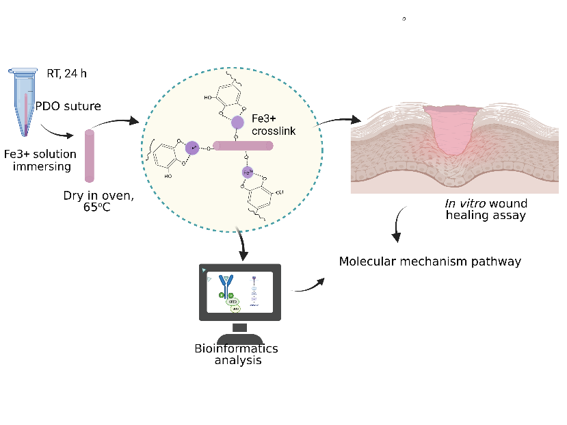
A surgical suture is a vital medical device to uphold wounded tissue during surgery. However, the application to improve tissue healing must still be developed. This study offered to solve this problem by incorporating tannic acid (TA) into polydioxanone (PDS) sutures. The PDS suture was immersed in TA complexing with iron (III) solutions in pH 8.0 solution. The engineered suture was successful, as revealed by the OH peak of TA in FTIR spectra. Furthermore, it was confirmed by TA particle appearance under Scanning Electron Microscopy. Contact angle measurement showed better wettability in biological mimicking solutions, such as water, serum, and saline solution. Thereby, it improved in vitro wound healing activity. Moreover, our bioinformatics data showed that TA enhanced the wound healing process by addressing multiple targets in the early phase of inflammation. For instance, it induced platelets to reduce bleeding at the early wound healing stage, activated T cells at the inflammation stage, accelerated epidermis cell proliferation, and activated fibroblast as a key player in wound healing. Due to multiple wound healing targets, our suture system was expected to be emphasized in clinical applications.
HIGHLIGHTS
- The engineered surgical suture was fabricated by modifying the surface of surgical suture with tannic acid (TA) through iron (III) complex formation in basic condition
- The modified PDS suture had a high wettability degree of water, saline, and serum, representing the biological fluid, which was expected to have better biocompatibility
- Based on bioinformatics analysis, TA revealed multiple targets to stop bleeding by activating platelet through FYN, CD4, and CD8 T cells, induced epithelial cells proliferation through Epidermal Growth Factor Receptor (EGFR), and activated many transcription and migration factors of fibroblast
GRAPHICAL ABSTRACT
Downloads
References
C Dennis, S Sethu, S Nayak, L Mohan, Y Morsi and G Manivasagam. Suture materials - current and emerging trends. J. Biomed. Mater. Res. 2016; 104, 1544-59.
C Yag-Howard. Sutures, needles, and tissue adhesives: A review for dermatologic surgery. Dermatol. Surg. 2014; 40, 2-15.
JC Kim, YK Lee, BS Lim, SH Rhee and HC Yang. Comparison of tensile and knot security properties of surgical sutures. J. Mater. Sci. Mater. Med. 2007; 18, 2363-9.
CKS Pillai and CP Sharma. Review paper: Absorbable polymeric surgical sutures: Chemistry, production, properties, biodegradability, and performance. J. Biomater. Appl. 2010; 25, 291-366.
KR Kunduru, A Basu and AJ Domb. Biodegradable polymers: Medical applications. Encyclopedia Polymer Sci. Tech. 2016, https://doi.org/10.1002/0471440264.pst027.pub2.
RE Abhari, JA Martins, HL Morris, PA Mouthuy and A Carr. Synthetic sutures: Clinical evaluation and future developments. J. Biomater. Appl. 2017; 32, 410-21.
B Joseph, A George, S Gopi, N Kalarikkal and S Thomas. Polymer sutures for simultaneous wound healing and drug delivery - a review. Int. J. Pharm. 2017; 524, 454-66.
JE Lee, Park, M Park, MH Kim, CG Park, SH Lee, SY Choi, BH Kim, HJ Park, JH Park, CY Heo and YB Choy. Surgical suture assembled with polymeric drug-delivery sheet for sustained, local pain relief. Acta Biomaterialia 2013; 9, 8318-27.
AL Gallo, F Paladini, A Romano, T Verri, A Quattrini, A Sannino and M Pollini. Efficacy of silver coated surgical sutures on bacterial contamination, cellular response and wound healing. Mater. Sci. Eng. C 2016; 69, 884-93.
RS Camenzind, TO Tondelli, T Götschi, C Holenstein and JG Snedeker. Can genipin-coated sutures deliver a collagen crosslinking agent to improve suture pullout in degenerated tendon? An Ex Vivo animal study. Clin. Orthop. Relat. Res. 2018; 476, 1104-12.
B Pasternak, A Missios, A Askendal, P Tengvall and P Aspenberg. Doxycycline-coated sutures improve the suture-holding capacity of the rat Achilles tendon. Acta Orthop. 2007; 78, 680-6.
HW Walling, DR Christensen, CJ Arpey and DC Whitaker. Surgical pearl: Lubrication of polyglactin suture with antibiotic ointment. J. Am. Acad. Dermatol. 2005; 52, 136-7.
MG Onesti, S Carella and N Scuderi. Effectiveness of antimicrobial-coated sutures for the prevention of surgical site infection: A review of the literature. Eur. Rev. Med. Pharmacol. Sci. 2018; 22, 5729-39.
YY Jo, H Kweon, DW Kim, MK Kim, SG Kim, JY Kim, WS Chae, SP Hong, YH Park, SY Lee and JY Choi. Accelerated biodegradation of silk sutures through matrix metalloproteinase activation by incorporating 4-hexylresorcinol. Sci. Rep. 2017; 7, 42441.
A Setiawati, D Jang, D Cho, S Cho, H Jeong, S Park, J Gwak, SR Ryu, WH Jung, BG Ju, KH Jung, OS Kwon and K Shin. An accelerated wound-healing surgical suture engineered with an extracellular matrix. Adv. Healthc. Mater. 2021; 10, 2001686.
M Xue and CJ Jackson. Extracellular matrix reorganization during wound healing and its impact on abnormal scarring. Adv. Wound Care 2015; 4, 119-36.
JK Ho and BM Hantash. The principles of wound healing. Expet. Rev. Dermatol. 2013; 8, 639-58.
K Wu, M Fu, Y Zhao, E Gerhard, Y Li, J Yang and J Guo. Anti-oxidant anti-inflammatory and antibacterial tannin-crosslinked citrate-based mussel-inspired bioadhesives facilitate scarless wound healing. Bioactive Mater. 2023; 20, 93-110.
Y Chen, L Tian, F Yang, W Tong, R Jia, Y Zou, L Yin, L Li, C He, X Liang, G Ye, C Lv, X Song and Z Yin. Tannic acid accelerates cutaneous wound healing in rats via activation of the ERK 1/2 signaling pathways. Adv. Wound Care 2019; 8, 341-54.
X Lv, L Wang, J Fu, Y Li and L Yu. A one-step tannic acid coating to improve cell adhesion and proliferation on polydimethylsiloxane. New J. Chem. 2020; 44, 15140-7.
F Sun, Y Bu, Y Chen, F Yang, J Yu and D Wu. An injectable and instant self-healing medical adhesive for wound sealing. ACS Appl. Mater. Interfac. 2020; 12, 9132-40.
C Hou, Y Wang, H Zhu and H Wei. Construction of enzyme immobilization system through metal-polyphenol assisted Fe3O4/chitosan hybrid microcapsules. Chem. Eng. J. 2016; 283, 397-403.
DG Barrett, TS Sileika and PB Messersmith. Molecular diversity in phenolic and polyphenolic precursors of tannin-inspired nanocoatings. Chem. Comm. 2014; 50, 7265-8.
P Networks, J Guo, Y Ping, H Ejima, K Alt, M Meissner, JJ Richardson, Y Yan, K Peter, D Elverfeldt, CE Hagemeyer and F Caruso. Engineering multifunctional capsules through the assembly of metal- phenolic networks. Angew. Chem. Int. Ed. Engl. 2014; 53, 5546-51.
H Ejima, JJ Richardson, K Liang, JP Best, MP Van Koeverden, GK Such, J Cui and F Caruso. One-step assembly of coordination complexes for versatile film and particle engineering. Science 2013; 341, 154-7.
AL Tajirian and DJ Goldberg. A review of sutures and other skin closure materials. J. Cosmet. Laser Ther. 2010; 12, 296-302.
KA Martins, AA Lach, HL Morris, AJ Carr and PA Mouthuy. Polydioxanone implants: A systematic review on safety and performance in the patients. J. Biomater. Appl. 2019; 34, 902-16.
N Goonoo, R Jeetah, A Bhaw-Luximon and D Jhurry. Polydioxanone-based biomaterials for tissue engineering and drug/gene delivery applications. Eur. J. Pharm. Biopharm. 2015; 97, 317-91.
P Shannon, A Markiel, O Ozier, NS Baliga, JT Wang, D Ramage, N Amin, B Schwikowski and T Ideker. Cytoscape: A software environment for integrated models of biomolecular interaction networks. Genome Res. 2003; 13, 2498-504.
D Szklarczyk, A Franceschini, S Wyder, K Forslund, D Heller, J Huerta-cepas, M Simonovic, A Roth, A Santos, KP Tsafou, M Kuhn, P Bork, LJ Jensen and CV Mering. STRING v10: Protein - protein interaction networks, integrated over the tree of life. Nucleic Acids Res. 2015; 43, D447-D452.
C Chia-Hao, C Shu-Hwa, W Hsin-Hung, H Chin-Wen, K Ming-Tat and L Chung-Yen. CytoHubba: Identifying hub objects and sub-networks from complex interactome. BMC Syst. Biol. 2014; 8, S11.
T Hhtamäki, X Tian, JT Korhonen and RHA Ras. Surface-wetting characterization using contact-angle measurements. Nat. Protocol. 2018; 13, 1521-38.
C McKenna. Simple and low-cost contact angle measurements using a smartphone with a PDMS-Lens. Royal Society of Chemistry, London, 2016.
D Pattarayan, A Sivanantham, R Bethunaickan, R Palanichamy and S Rajasekaran. Tannic acid modulates fibroblast proliferation and differentiation in response to pro-fibrotic stimuli. J. Cell. Biochem. 2018; 119, 6732-42.
N Dumitrascu and C Borcia. Determining the contact angle between liquids and cylindrical surfaces. J. Colloid Interface Sci. 2006; 294, 418-22.
KL Menzies and L Jones. The impact of contact angle on the biocompatibility of biomaterials. Optom. Vis. Sci. 2010; 87, 387-99.
F Jimenez, TF Mitts, K Liu, Y Wang and A Hinek. Ellagic and tannic acids protect newly synthesized elastic fibers from premature enzymatic degradation in dermal fibroblast cultures. J. Investig. Dermatol. 2006; 126, 1272-80.
CAP Cass and KJL Burg. Tannic acid cross-linked collagen scaffolds and their anti-cancer potential in a tissue engineered breast implant. J. Biomater. Sci. Polymer Ed. 2012; 23, 281-98.
HB Nygaard, CHV Dyck and SM Strittmatter. Fyn kinase inhibition as a novel therapy for Alzheimer’s disease. Alzheimer’s Res. Ther. 2014; 6, 8.
JA Dudakov, AM Hanash and MRMVD Brink. Interleukin-22: Immunobiology and pathology. Annu. Rev. Immunol. 2015; 33, 747-85.
G Pickert, C Neufert, M Leppkes, Y Zheng, N Wittkopf, M Warntjen, H Lehr, S Hirth, B Weigmann, S Wirtz, W Ouyang, MF Neurath and C Becker. STAT3 links IL-22 signaling in intestinal epithelial cells to mucosal wound healing. J. Exp. Med. 2009; 206, 1465-72.
HM Mcgee, BA Schmidt, CJ Booth, GD Yancopoulos, DM Valenzuela, AJ Murphy, S Stevens, RA Flavell and V Horsley. IL-22 promotes fibroblast-mediated wound repair in the skin. J. Investig. Dermatol. 2012; 133, 1321-9.
K Raziyeva, Y Kim, Z Zharkinbekov, K Kassymbek, S Jimi and A Saparov. Immunology of acute and chronic wound healing. Biomoluecules 2021; 11, 700.
W Li, D Sahu and F Tsen. Secreted heat shock protein-90 (Hsp90) in wound healing and cancer. Biochim. Biophys. Acta Mol. Cell Res. 2012; 1823, 730-41.
J Larouche, S Sheoran and K Maruyama. Immune regulation of skin wound healing: mechanisms and novel therapeutic targets. Adv. Wound Care 2018; 7, 209-31.
S Nakerakanti and M Trojanowska. The role of TGF-receptors in fibrosis. Open Rheumatol. J. 2012; 6, 156-62.
M Deng, W Chen, A Takatori, Z Peng, L Zhang, M Mongan, R Parthasarathy, M Sartor, M Miller, J Yang, B Su, WW Kao and Y Xia. A role for the mitogen-activated protein kinase kinase kinase 1 in epithelial wound healing. Mol. Biol. Cell 2006; 17, 3446-55.
KB Reddy, DM Smith and EF Plow. Analysis of Fyn function in hemostasis and α IIb β 3- integrin signaling. J. Cell Sci. 2008; 121, 1641-8.
EM Golebiewska and AW Poole. Platelet secretion: From haemostasis to wound healing and beyond. Blood Rev. 2015; 29, 153-62.
A Allam, M Yakou, L Pang, M Ernst, J Huynh, CM Marzocchi-machado and P Trono. Exploiting the STAT3 nexus in cancer-associated fibroblasts to improve cancer therapy. Front. Immunol. 2021; 12, 767939.
MM Kasembeli, U Bharadwaj, P Robinson and DJ Tweardy. Contribution of STAT3 to inflammatory and fibrotic diseases and prospects for its targeting for treatment. Int. J. Mol. Sci. 2018; 19, 2299.
Y Matsubayashi, M Ebisuya, S Honjoh and E Nishida. ERK activation propagates in epithelial cell sheets and regulates their migration during wound healing. Curr. Biol. 2004; 14, 731-5.
L Vaidyanathan. Growth factors in wound healing - a review. Biomed. Pharmacol. J. 2021; 14, 1469-80.
Y Nakamura, C Sotozono and S Kinoshita. The epidermal growth factor receptor (EGFR): Role in corneal wound healing and homeostasis. Exp. Eye Res. 2001; 2, 511-7.
F Barone, S Nayar, J Campos, T Cloake, DR Withers, KM Toellner, Y Zhang, L Fouser, B Fisher, S Bowman, J Rangel-Moreno, MDLL Garcia-Hernandez, TD Randall, D Lucchesi, M Bombardieri, C Pitzalis, SA Luther and CD Buckley. IL-22 regulates lymphoid chemokine production and assembly of tertiary lymphoid organs. Proc. Nat. Acad. Sci. Unit. States Am. 2015; 112, 11024-9.
J Rossy, DJ Williamson and K Gaus. How does the kinase Lck phosphorylate the T cell receptor ? Spatial organization as a regulatory mechanism. Front. Immunol. 2012; 3, 167.
D Scieglinska, Z Krawczyk, DR Sojka and A Gogler-Pigłowska. Heat shock proteins in the physiology and pathophysiology of epidermal keratinocytes. Cell Stress Chaperones 2019; 24, 1027-44.
C Wiegand, U Hipler, P Elsner and J Tittelbach. Keratinocyte and fibroblast wound healing in vitro is repressed by non-optimal conditions but the reparative potential can be improved by water-filtered infrared A. Biomedicines 2021; 9, 1802.
Downloads
Published
How to Cite
Issue
Section
License
Copyright (c) 2023 Walailak University

This work is licensed under a Creative Commons Attribution-NonCommercial-NoDerivatives 4.0 International License.


