
Folic Acid Attenuates MSG-Induced Teratogenicity during A 2-Month Pregnancy by Preventing Neural Crest Cell Destruction and Malformation in Chick Embryo Models
DOI:
https://doi.org/10.48048/tis.2023.6656Keywords:
Monosodium glutamate, Chick embryo, Birth defect, Folic acid, Neural crest cellsAbstract
Monosodium glutamate (MSG), commonly used as a food enhancer, has been reported to have teratogenicity during the first 3 days of development. Furthermore, the neural crest cells (NCCs) are crucial for embryonic development during organogenesis. The present study aimed to investigate the treatment effect of folic acid (FA) on MSG-induced teratogenicity, focusing on the toxicity and teratogenic effects on somatic and neural crest cells in chick embryos as models. Six hundred and fifty fertilized eggs were divided into control, FA, MSG, and MSG with FA groups. The chemicals were administered, and the results were investigated after 3 days of incubation. The morphology and histology were studied using stereomicroscopy and hematoxylin and eosin staining, respectively. The NCC population was confirmed by the presence of HNK-1 using immunohistochemistry. The finding showed that the MSG at 2 mg/kg of egg weight induced retardation, tissue malformation, craniofacial, and heart defects, whereas the FA alleviated those adverse effects and reduced the MSG-induced NCCs destruction in the eyes, heart, stomach, and nerves. In conclusion, although MSG harms embryos, FA effectively diminished its teratogenicity in the chick embryo model. These experimental protocols are beneficial for teratogenic studies on preventing birth defects that are harmful to the embryo.
HIGHLIGHTS
- The 4 mg/kg egg weight of monosodium glutamate exposure leads to chick embryo lethality
- Folic acid treatment is effective at 2 mg/kg of monosodium glutamate exposure
- Folic acid is essential for supervised neural crest cell functions following monosodium glutamate-induced teratogenesis
- These experimental protocols are beneficial for teratogenic studies
GRAPHICAL ABSTRACT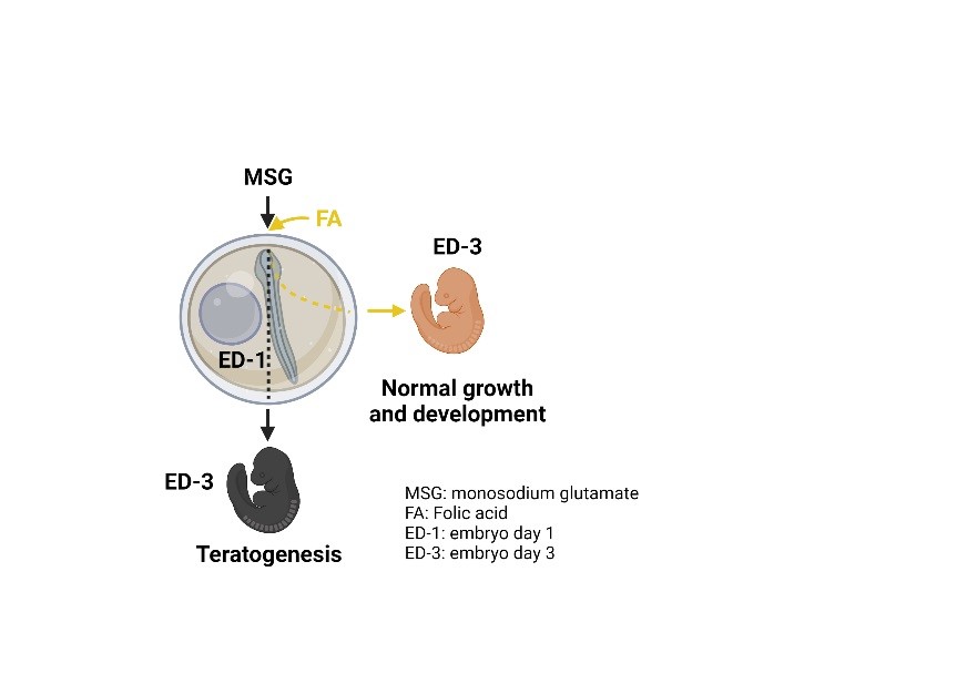
Downloads
References
LE Mitchell. Maternal effect genes: Update and review of evidence for a link with birth defects. HGG Adv. 2022; 3, 100067.
A Christianson, CP Howson and B Modell. March of dimes: Global report on birth defects, the hidden toll of dying and disabled children. March of Dimes Birth Defects Foundation. Virginia, 2005.
Y Zhou, X Mao, H Zhou, L Wang, Z Qin, Z Cai and B Yu. Birth defects data from population-based birth defects surveillance system in a district of Southern Jiangsu, China, 2014-2018. Front. Publ. Health 2020; 8, 378.
ML Feldkamp, JC Carey, JL Byrne, S Krikov and LD Botto. Etiology and clinical presentation of birth defects: Population based study. BMJ 2017; 357, j2249.
GE Wachholz, BD Rengel, N Vargesson and LR Fraga. From the farm to the lab: How chicken embryos contribute to the field of teratology. Front. Genet. 2021; 12, 666726.
D Zosen, MG Hadera, JS Lumor, JM Andersen and RE Paulsen. Chicken embryo as animal model to study drug distribution to the developing brain. J. Pharmacol. Toxicol. Meth. 2021; 112, 107105.
MN Vergara and MV Canto-Soler. Rediscovering the chick embryo as a model to study retinal development. Neural Dev. 2012; 7, 22.
RF Gasser, RJ Cork, BJ Stillwell and DT McWilliams. Rebirth of human embryology. Dev. Dynam. 2014; 243, 621-8.
AV Hoffbrand and DG Weir. The history of folic acid. Br. J. Haematol. 2001; 113, 579-89.
HK Mitchell, EE Snell and RJ Williams. The concentration of “folic acid”. J. Am. Chem. Soc. 1941; 63, 2284.
IH Rosenberg. A history of the isolation and identification of folic acid (folate). Ann. Nutr. Metabol. 2012; 61, 231-5.
JG Donnelly. Folic acid. Crit. Rev. Clin. Lab. Sci. 2001; 38, 183-223.
K Sato. Why is folate effective in preventing neural tube closure defects? Med Hypotheses 2020; 134, 109429.
D Rothman. Folic acid in pregnancy. Am. J. Obstet. Gynecol. 1970; 108, 149.
BDL Fournière, F Dhombres, P Maurice, SD Foucaud, P Lallemant, M Zérah, L Guilbaud and J Jean-Marie. Prevention of neural tube defects by folic acid supplementation: A national population-based study. Nutrients 2020; 12, 3170.
CD Dolin, AL Deierlein and MI Evans. Folic acid supplementation to prevent recurrent neural tube defects: 4 milligrams is too much. Fetal Diagn. Ther. 2018; 44, 161-5.
KM Appaiah. Chapter 13 - monosodium glutamate in foods and its biological effects. Ensuring Global Food Saf. 2010; 2010, 217-26.
K Ganesan, K Sukalingam, K Balamurali, SRBS Alaudeen, K Ponnusamy, IA Ariffin and SB Gani. A studies on monosodium L-Glutamate toxicity in animal models-a review. Int. J. Pharmaceut. Chem. Biol. Sci. 2013; 3, 1257-68.
M Freeman. Reconsidering the effects of monosodium glutamate: A literature review. J. Am. Assoc. Nurse Pract. 2006; 18, 482-6.
Z Kazmi, I Fatima, S Perveen and SS Malik. Monosodium glutamate: Review on clinical reports. Int. J. Food Properties 2017; 20, 1807-15.
K Niaz, E Zaplatic and J Spoor. Extensive use of monosodium glutamate: A threat to public health? EXCLI J. 2018; 17, 273.
K Beyreuther, HK Biesalski, JD Fernstrom, P Grimm, WP Hammes, U Heinemann, O Kempski, P Stehle, H Steinhart and R Walker. Consensus meeting: Monosodium glutamate-an update. Eur. J. Clin. Nutr. 2007; 61, 304-13.
J Roongruangchai, Y Viravud, V Plakornkul, K Sripaoraya, W Boonmark and K Roongruangchai. The teratogenic effects of monosodium glutamate (MSG) on the development of chick embryos. Siriraj Med. J. 2018; 70, 514-22.
F Al-Qudsi and A Al-Jahdali. Effect of monosodium glutamate on chick embryo development. J. Am. Sci. 2012; 8, 499-509.
J Casale and AO Giwa. Embryology, branchial arches. StatPearls, Florida, 2022.
R Hunt and PN Hunt. The role of cell mixing in branchial arch development. Mech. Dev. 2003; 120, 769-90.
MB Carbonell, RF Bayona, Z Garavito-Aguilar, BC Parada, GH Arboleda and C Infante-Contreras. Hey1 gene expression patterns during the development of branchial arches and facial prominences. Rev. MVZ Córdoba 2018; 23, 6813-25.
MR Passos‐Bueno, CC Ornelas and RD Fanganiello. Syndromes of the first and second pharyngeal arches: A review. Am. J. Med. Genet. 2009; 149, 1853-9.
JM Johnson, G Moonis, GE Green, R Carmody and HN Burbank. Syndromes of the first and second branchial arches, part 1: Embryology and characteristic defects. Am. J. Neuroradiol. 2011; 32, 14-9.
MED Bellard, Y Rao and M Bronner-Fraser. Dual function of Slit2 in repulsion and enhanced migration of trunk, but not vagal, neural crest cells. Int. J. Cell Biol. 2003; 162, 269-79.
ME Bronner and MN LeDouarin. Development and evolution of the neural crest: An overview. Dev. Biol. 2012; 366, 2-9.
P Noisa and T Raivio. Neural crest cells: From developmental biology to clinical interventions. Birth Defect. Res. C Embryo Today Rev. 2014; 102, 263-74.
TL Creazzo, RE Godt, L Leatherbury, SJ Conway and ML Kirby. Role of cardiac neural crest cells in cardiovascular development. Annu. Rev. Physiol. 1998; 60, 267.
EM Siismets and NE Hatch. Cranial neural crest cells and their role in the pathogenesis of craniofacial anomalies and coronal craniosynostosis. J. Dev. Biol. 2020; 8, 18.
S Cerrizuela, GA Vega‐Lopez and MJ Aybar. The role of teratogens in neural crest development. Birth Defect. Res. 2020; 112, 584-632.
JP Saint-Jeannet, P Blader, LA Taneyhill. Cranial placodes and neural crest interactions in craniofacial development. Front. Physiol. 2021; 12, 681397.
Y Shi, J Li, C Chen, M Gong, Y Chen, Y Liu, J Chen, T Li and W Song. 5-Mehtyltetrahydrofolate rescues alcohol-induced neural crest cell migration abnormalities. Mol. Brain 2014; 7, 67.
NAA Elnaga, M Sarhan and H Mansour. Teratogenicity of monosodium glutamate on the pregnant rats and their fetuses. Egypt. J. Hosp. Med. 2019; 74, 1737-47.
CP Chen. Syndromes, disorders and maternal risk factors associated with neural tube defects (V). Taiwanese J. Obstet. Gynecol. 2008; 47, 259-66.
GJ Mahler and JT Butcher. Cardiac developmental toxicity. Birth Defect. Res. C Embryo Today Rev. 2011; 93, 291-7.
AS Mahaliyana, MFA Fasmina, AMTB Alahakoon and GMGMM Wickrama. Toxicity effects of monosodium glutamate (MSG) on embryonic development of zebrafish (Danio rerio); a promising model to study excitotoxins. Int. J. Sci. Res. 2016; 6, 229-34.
TA Lynch and DE Abel. Teratogens and congenital heart disease. J. Diagn. Med. Sonography 2015; 31, 301-5.
J Tikkanen and OP Heinonen. Congenital heart disease in the offspring and maternal habits and home exposures during pregnancy. Teratology 1992; 46, 447-54.
E Gilbert-Barness. Teratogenic causes of malformations. Ann. Clin. Lab. Sci. 2010; 40, 99-114.
T Kuribayashi and WC Roberts. Tetralogy of fallot, truncus arteriosus, abnormal myocardial architecture and anomalies of the aortic arch system induced by bis-diamine in rat fetuses. J. Am. Coll. Cardiol. 1993; 21, 768-76.
A Sharma. Monosodium glutamate-induced oxidative kidney damage and possible mechanisms: A mini-review. J. Biomed. Sci. 2015; 22, 1-6.
KR George, NG Shibija and NA Malini. Monosodium glutamate (MSG) induced developmental dysfunction in female albino rats (Rattus norvegicus). Bioscan 2013; 8, 73-6.
FA Eid, NA Abu Elnaga, M Sarhan and H Mansour. Effect of monosodium glutamate on liver of pregnant rats and their fetuses (Histological and histochemical studies). Egypt. J. Hosp. Med. 2018; 73, 8091-8.
CE Butterworth and A Bendich. Folic acid and the prevention of birth defects. Annu. Rev. Nutr. 1996; 16, 73-97.
RA Swain and LS Clair. The role of folic acid in deficiency states and prevention of disease. J. Fam. Pract. 1997; 44, 138-44.
J Safi, L Joyeux and GE Chalouhi. Periconceptional folate deficiency and implications in neural tube defects. J. Pregnancy 2012; 2012, 295083.
AO Lucas, BJ Stoll and JR Bale. Improving birth outcomes: Meeting the challenge in the developing world. National Academies Press, Washington, 2003.
R Zhao, RG Russell, Y Wang, L Liu, F Gao, B Kneitz, W Edelmann and ID Goldman. Rescue of embryonic lethality in reduced folate carrier-deficient mice by maternal folic acid supplementation reveals early neonatal failure of hematopoietic organs. J. Biol. Chem. 2001; 276, 10224-8.
YI Goh and G Koren. Folic acid in pregnancy and fetal outcomes. J. Obstet. Gynaecol. 2008; 28, 3-13.
EO Farombi and OO Onyema. Monosodium glutamate-induced oxidative damage and genotoxicity in the rat: modulatory role of vitamin C, vitamin E and quercetin. Hum. Exp. Toxicol. 2006; 25, 251-9.
DAE Hassan, MAA Alim, SMZ Sharkawi and S Nabil. Detection of cardiac tissues toxicity caused by monosodium glutamate and the protective role of vitamin c by immunohistochemical method, heart tissue oxidative stress biomarkers and cardiac dysfunction biomarkers. Egypt. J. Forensic Sci. Appl. Toxicol. 2020; 20, 13-21.
W Li, Y Ma, Z Li, X Lv, X Wang, D Zhou, S Luo, JX Wilson and G Huang. Folic acid decreases astrocyte apoptosis by preventing oxidative stress-induced telomere attrition. Int. J. Mol. Sci. 2019; 21, 62.
ML Kirby and KL Waldo. Role of neural crest in congenital heart disease. Circulation 1990; 82, 332-40.
M Maroto, R Reshef, AE Münsterberg, S Koester, M Goulding and AB Lassar. Ectopic Pax-3 activates MyoD and Myf-5 expression in embryonic mesoderm and neural tissue. Cell 1997; 89, 139-48.
S Tajbakhsh, D Rocancourt, G Cossu and M Buckingham. Redefining the genetic hierarchies controlling skeletal myogenesis: Pax-3 and Myf-5 act upstream of MyoD. Cell 1997; 89, 127-38.
AG Borycki, J Li, FUZI Jin, CP Emerson and JA Epstein. Pax3 functions in cell survival and in pax7 regulation. Development 1999; 126, 1665-74.
FG Barr, N Galili, J Holick, JA Biegel, G Rovera and BS Emanuel. Rearrangement of the PAX3 paired box gene in the paediatric solid tumour alveolar rhabdomyosarcoma. Nat. Genet. 1993; 3, 113-7.
N Galili, RJ Davis, WJ Fredericks, S Mukhopadhyay, FJ Rauscher, BS Emanuel, G Rovera and FG Barr. Fusion of a fork head domain gene to PAX3 in the solid tumour alveolar rhabdomyosarcoma. Nat. Genet. 1993; 5, 230-5.
DN Shapiro, JE Sublett, B Li, JR Downing and CW Naeve. Fusion of PAX3 to a member of the forkhead family of transcription factors in human alveolar rhabdomyosarcoma. Canc. Res. 1993; 53, 5108-12.
JA Epstein, J Li, D Lang, F Chen, CB Brown, F Jin, MM Lu, M Thomas, E Liu, A Wessels and CW Lo. Migration of cardiac neural crest cells in splotch embryos. Development 2000; 127, 1869-78.

Downloads
Published
How to Cite
Issue
Section
License
Copyright (c) 2023 Walailak University

This work is licensed under a Creative Commons Attribution-NonCommercial-NoDerivatives 4.0 International License.


