
Evaluation of The Hepatoprotective Effect of Plantago major Extract in A Rifampicin-Isoniazid Induced Hepatitis Rat Model
DOI:
https://doi.org/10.48048/tis.2023.6331Keywords:
Antituberculosis Drugs, Hepatoprotection, Histopathology, Isoniazid, MDA, Plantago major, Rifampicin, SGPTAbstract
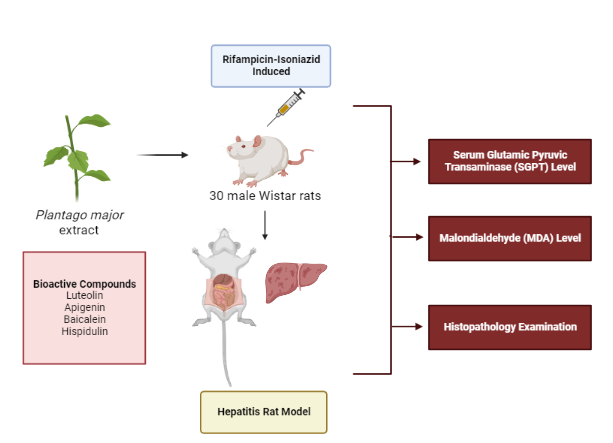
Tuberculosis (TB) is an ancient human disease caused by Mycobacterium tuberculosis which affects the lungs, making pulmonary disease the most common presentation. Herbal medicine began to be developed as hepatoprotection. The hepatoprotection effects of Plantago major extract on serum glutamic pyruvic transaminase (SGPT), liver tissue Malondialdehyde (MDA), and histopathological changes were evaluated in rifampicin and isoniazid-induced hepatitis rats. Thirty male Wistar rats were divided into 6 groups: Group I received 1 % PGA, with hepatitis induced in the remaining groups by rifampicin and isoniazid 50 mg/kg BW/day, group II was only given rifampicin and isoniazid, group III was given curcuma 10.8 mg/kg BW/day and groups IV (PM1), V (PM2), and VI (PM3) were given Plantago major extract at a dose of 20.3, 40.5, and 81 mg/kg BW/day, respectively. The rats were treated for 28 days. Administration of Plantago major extract (20.3 and 40.5 mg/kg BW/day) inhibited the elevation of serum SGPT and MDA levels, with less portal inflammation than the negative control group. The rats treated with the higher dose of 81 mg/kg BW/day had serum SGPT, MDA, and percentage portal inflammation equivalent to the negative control group. The Plantago major extract at a dose of 20.3 and 40.5 mg/kg BW can inhibit the elevated serum SGPT. Plantago major extract exerts dose-dependent hepatoprotection effects in a rifampicin-isoniazid induced hepatitis rat model by reducing elevated levels of SGPT, liver tissue MDA, as well as the percentage of portal inflammation.
HIGHLIGHTS
- To the knowledge of the authors, there has been no comprehensive work dedicated to antituberculosis drugs
- The administration of Plantago major extract showed hepatoprotector effects, as evidenced by the inhibition of elevated levels of SGPT, MDA, and a lower percentage of portal inflammation than the negative controls
- Plantago major extract can be used as herbal medicine that can be juxtaposed with the main drug in TB patients
GRAPHICAL ABSTRACT
Downloads
References
V Ramappa and GP Aithal. Hepatotoxicity related to anti-tuberculosis drugs: Mechanisms and management. J. Clin. Exp. Hepatol. 2013; 3, 37-49.
World Health Organization. Global tuberculosis report. World Health Organization, Geneva, Switzerland, 2019.
P Shang, Y Xia, F Liu, X Wang, Y Yuan, D Hu, D Tu, Y Chen, P Deng, S Cheng and L Zhou. Incidence, clinical features, and impact on anti-tuberculosis treatment of anti-tuberculosis drug induced liver injury (ATLI) in China. PLoS One 2011; 6, e21836.
SA Rasyid, Armayani, Yuniati and TMP Lio. Analysis of serum glutamic pyruvic transaminase and serum glutamic oxaloacetic transaminase levels in tuberculosis patients who are undergoing oat treatment in Kendari City General Hospital, Kota Kendari, Indonesia. Infect. Dis. Rep. 2020; 12, 8737.
RK TA, S Khan, P Sen and S Banerjee. A Study to detect liver enzyme dysfunction among patients on first line anti-tubercular drugs from RNTCP during the course of anti-TB treatment. J. Evol. Med. Dent. Sci. 2020; 9, 645-50.
MB Adom, M Taher, MF Mutalabisin, MS Amri, MB Kudos, MW Sulaiman, P Sengupta and D Susanti. Chemical constituents and medical benefits of Plantago major. Biomed. Pharmacother. 2017; 96, 348-60.
T Akbarzadeh, R Sabourian, M Saeedi, H Rezaeizadeh, M Khanavi and MR Ardekani. Liver tonics: Review of plants used in Iranian traditional medicine. Asian Pac. J. Trop. Biomed. 2015; 5, 170-81.
AH Eldesoky, RF Abdel‐Rahman, OK Ahmed, GA Soliman, AS Saeedan, HY Elzorba, AA Elansary and M Hattori. Antioxidant and hepatoprotective potential of Plantago major growing in Egypt and its major phenylethanoid glycoside, acteoside. J. Food Biochem. 2018; 42, e12567.
Y Najafian, SS Hamedi, MK Farshchi and Z Feyzabadi. Plantago major in Traditional Persian Medicine and modern phytotherapy: A narrative review. Electron. Physician. 2018; 10, 6390.
ZA Zaenah. Effect of luteolin and quercetin on thioacetamide induced hepatic fibrosis in rats. Int. J. Pharmacol. 2019; 15, 863-71.
J Khan, S Saraf and S Saraf. Preparation and evaluation of luteolin–phospholipid complex as an effective drug delivery tool against GalN/LPS induced liver damage. Pharm. Dev. Tech. 2016; 21, 475-86.
A Rašković, S Gigov, I Čapo, M Paut Kusturica, B Milijašević, S Kojić-Damjanov and N Martić. Antioxidative and protective actions of apigenin in a paracetamol-induced hepatotoxicity rat model. Eur. J. Drug Metab. Pharmacokinet. 2017; 42, 849-56.
B Salehi, A Venditti, M Sharifi-Rad, D Kręgiel, J Sharifi-Rad, A Durazzo, M Lucarini, A Santini, EB Souto, E Novellino and H Antolak. The therapeutic potential of apigenin. Int. J. Mol. Sci. 2019; 20, 1305.
HL Huang, YJ Wang, QY Zhang, B Liu, FY Wang, JJ Li and RZ Zhu. Hepatoprotective effects of baicalein against CCl4-induced acute liver injury in mice. World J. Gastroenterol. 2012; 18, 6605.
HC Zhou, H Wang, K Shi, JM Li, Y Zong and R Du. Hepatoprotective effect of baicalein against acetaminophen-induced acute liver injury in mice. Molecules 2018; 24, 131.
K Patel and DK Patel. Medicinal importance, pharmacological activities, and analytical aspects of hispidulin: A concise report. J. Tradit. Complement Med. 2017; 7, 360-6.
F Hussan, RH Basah, MR Yusof, NA Kamaruddin and F Othman. Plantago major treatment enhanced innate antioxidant activity in experimental acetaminophen toxicity. Asian Pac. J. Trop. Biomed. 2015; 5, 728-32.
IK Mohamed, MA Osama, M Samiha and EM Zahrat. Biochemical studies on Plantago major L. and Cyamopsis tetragonoloba L. Int. J. Biodivers. Conserv. 2011; 3, 83-91.
PY Kwo, SM Cohen and JK Lim. ACG clinical guideline: Evaluation of abnormal liver chemistries. Am. J. Gastroenterol. 2017; 112, 18-35.
MR Pincus, PM Tierno, E Gleeson, WB Bowne and MH Bluth. Evaluation of liver function. In: R McPherson and M Pincus (Eds.). Henry’s clinical diagnosis and management by laboratory methods. 23rd ed. Elsevier, Amsterdam, Netherlands, 2017, p. 295-6.
Z Chen, Y Yang, S Mi, Q Fan, X Sun, B Deng, G Wu, Y Li, Q Zhou and Z Ruan. Hepatoprotective effect of chlorogenic acid against chronic liver injury in inflammatory rats. J. Funct. Foods. 2019; 62, 103540.
N Azarmehr, P Afshar, M Moradi, H Sadeghi, H Sadeghi, B Alipoor, B Khalvati, Z Barmoudeh, K Abbaszadeh-Goudarzi and AH Doustimotlagh. Hepatoprotective and antioxidant activity of watercress extract on acetaminophen-induced hepatotoxicity in rats. Heliyon 2019; 5, e02072.
SY Tiya, CR Sewani-Rusike and T Taderera. Hepatoprotective effects of Fadogia ancylantha (Makoni Tea) on ethanol-induced liver damage in Wistar rats. J. Biol. Act. Prod. Nat. 2019; 9, 352-63.
G Jiang, C Sun, X Wang, J Mei, C Li, H Zhan, Y Liao, Y Zhu and J Mao. Hepatoprotective mechanism of Silybum marianum on nonalcoholic fatty liver disease based on network pharmacology and experimental verification. Bioengineered 2022; 13, 5216-35.
MB Kadiiska, BC Gladen, DD Baird, D Germolec, LB Graham, CE Parker, A Nyska, JT Wachsman, BN Ames, S Basu and N Brot. Biomarkers of oxidative stress study II are oxidation products of lipids, proteins, and DNA markers of CCl4 poisoning. Free Radic. Biol. Med. 2005; 38, 698-710.
P Thérond, D Bonnefont-Rousselot, A Davit-Spraul, M Conti and A Legrand. Biomarkers of oxidative stress: An analytical approach. Curr. Opin. Clin. Nutr. Metab. Care 2000; 3, 373-84.
RA McPherson and MR Pincus. Henry’s clinical diagnosis and management by laboratory methods. 24th ed. Elsevier, Amsterdam, Netherlands, 2021.
M Krishna. Patterns of necrosis in liver disease. Clin. Liver Dis. 2017; 10, 53.
R Ramachandran and S Kakar. Histological patterns in drug-induced liver disease. J. Clin. Pathol. 2009; 62, 481-92.
MA Steele, RF Burk and RM DesPrez. Toxic hepatitis with isoniazid and rifampicin: A meta-analysis. Chest 1991; 99, 465-71.
I Shimizu, N Shimamoto, K Saiki, M Furujo and K Osawa. Lipid peroxidation in hepatic fibrosis. In: A Catala (Ed.). Lipid Peroxidation. IntechOpen, London, 2012, p. 483-92.
JJ Saukkonen, DL Cohn, RM Jasmer, S Schenker, JA Jereb, CM Nolan, CA Peloquin, FM Gordin, D Nunes, DB Strader and J Bernardo. An official ATS statement: Hepatotoxicity of antituberculosis therapy. Am. J. Respir. Crit. Care Med. 2006; 174, 935-52.
MH Farzaei, M Zobeiri, F Parvizi, FF El-Senduny, I Marmouzi, E Coy-Barrera, R Naseri, SM Nabavi, R Rahimi and M Abdollahi. Curcumin in liver diseases: A systematic review of the cellular mechanisms of oxidative stress and clinical perspective. Nutrients 2018; 10, 855.
Badan Pengawas Obat dan Makanan RI. Acuan Sediaan Herbal (in Indonesian). Vol VI. Badan Pengawas Obat dan Makanan RI, Jakarta, Indonesia, 2011.
OO Adewale, SS Samuel, M Manubolu and K Pathakoti. Curcumin protects sodium nitrite-induced hepatotoxicity in Wistar rats. Toxicol. Rep. 2019; 6, 1006-11.
R Singh and P Sharma. Hepatoprotective effect of curcumin on lindane-induced oxidative stress in male wistar rats. Toxicol. Int. 2011; 18, 124.
S Ganiger, HN Malleshappa, H Krishnappa, G Rajashekhar, VR Rao and F Sullivan. A two-generation reproductive toxicity study with curcumin, turmeric yellow, in Wistar rats. Food Chem. Toxicol. 2007; 45, 64-9.
National Library of Medicine. Clinical development plan: Curcumin. J. Cell Biochem. Suppl. 1996; 26, 72-85.
JL Guil, I Rodríguez-Garcí and E Torija. Nutritional and toxic factors in selected wild edible plants. Plant foods for human nutrition. 1997; 51, 99-107.
E Bozcali, Ö Süzer, HN Gürsoy, P Atukeren and KM Gümüstas. Effects of erucic acid supplemented feeding on chronic doxorubucin toxicity in rats. Int. J. Clin. Exp. Med. 2009; 2, 337.
DD Rio, AJ Stewart and N Pellegrini. A review of recent studies on malondialdehyde as toxic molecule and biological marker of oxidative stress. Nutr. Metab. Cardiovasc. Dis. 2005; 15, 316-28.
DH Kim, SJ Kwack, KS Yoon, JS Choi and BM Lee. 4-Hydroxynonenal: A superior oxidative biomarker compared to malondialdehyde, and carbonyl content induced by carbon tetrachloride in rats. J. Toxicol. Environ. Health A 2015; 78, 1051-62.
Downloads
Published
How to Cite
Issue
Section
License
Copyright (c) 2023 Walailak University

This work is licensed under a Creative Commons Attribution-NonCommercial-NoDerivatives 4.0 International License.


