
Effect of Vitamin D3 Treatment on Genes Expression of Corticotrophin Releasing Hormone (CRH), CRH Receptor 1 (CRH-R1) and Connexin-43 (CON-43) in PHM1-41 Cell Line that Induced by Hypoxia
DOI:
https://doi.org/10.48048/tis.2022.6236Keywords:
Corticotrophin releasing hormone, CRH receptor 1, Connexin-43, Oxidative stress, Vitamin D3Abstract
Introduction: Hypothalamic-Pituitary-Adrenal (HPA) axis activity is one of pathophysiologic mechanism that caused preterm labor. Biologic maternal stress, for instance hypoxia condition, is one of the causes that can trigger preterm birth occasion through the activation of HPA axis. It increases Corticotrophin Releasing Hormone (CRH), CRH receptor 1 (CRH-R1), and Connexin-43 (CON-43) as the trigger of the contraction process. Vitamin D as a source of Ca2+ ion is needed for myometrium smooth muscle’s concentrations and relaxation mechanism. The aim of this study was to determine the effect of vitamin D3 in PHM1-41 cell line. Materials and Methods: The human smooth muscle uterine myometrium cell line PHM1-41 as an in vitro model experimental subject, treated by hypoxia oxidative stress condition and added by vitamin D3 (5, 10, 50 and 150 nM). The dichlorodihydrofluorescein diacetate (DCFDA) fluorescent was used to measure the level of intracellular reactive oxygen species (ROS). In addition, RNA of treated PHM1-41cells was isolated for analyzing gene expressions such as CRH, CRH-R1, and CON-43 as a profile of contractility regulation. Results: ROS level effectively decrease in the cells that treated by 150 nM vitamin D3 group compared to the control hypoxia cell group (7.16 ± 0.23 and 19.49 ± 1.76, respectively). Expression of CRH, CRH-R1, and CON-43 genes are also decrease by treated with 150 nM vitamin D3 to the cells. Pearson (parametric) correlation analysis evidenced a negative correlation between the vitamin D3 additional treated to the lower of ROS level, CRH, CRH-R1, and CON-43 genes expression on PHM1-41 cell line that induced by oxidative stress condition. Conclusion: The concentration of 150 nM vitamin D3 was a prominent potency to prevent the incidence of preterm labor.
HIGHLIGHTS
- Hypothalamic-Pituitary-Adrenal (HPA) axis is one of the causes of preterm birth that marked by high level of intracellular ROS leads to increasing of Corticotrophin Releasing Hormone (CRH) which has significant role in aterm and preterm labor
- PHMI-41 cells can describe the Corticotrophin Releasing Hormone (CRH), CRH receptor 1 (CRH-R1), and Connexin-43 (CON-43) genes expression level so it was used as model of human pregnancy condition
- Vitamin D3 is one of antioxidant agent that can decrease ROS level also CRH, CRH-R1, and CON-43 genes expression on PHM1-41 cell especially at doses of 150 nM which shown a prominent potency to prevent the preterm labor incidence
GRAPHICAL ABSTRACT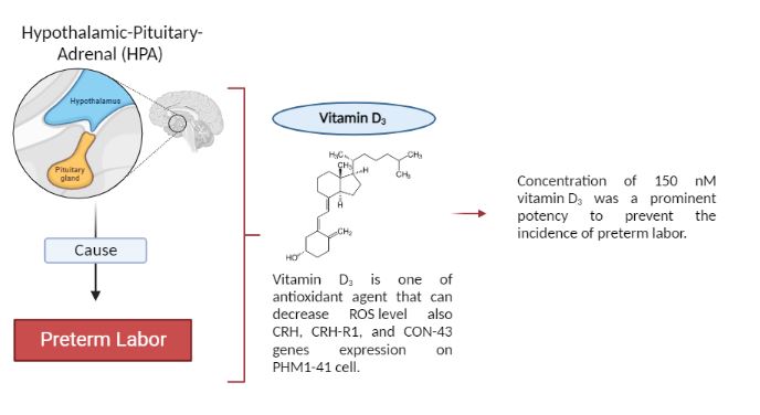
Downloads
References
JM Fettweis, MG Serrano, JP Brooks, DJ Edwards, PH Girerd, HI Parikh, B Huang, TJ Arodz, L Edupuganti, AL Glascock and J Xu. The vaginal microbiome and preterm birth. Nat. Med. 2019; 25, 1012-21.
S Chawanpaiboon, JP Vogel, AB Moller, P Lumbiganon, M Petzold, D Hogan, S Landoulsi, N Jampathong, K Kongwattanakul, M Laopaiboon and C Lewis. Global, regional, and national estimates of levels of preterm birth in 2014: A systematic review and modelling analysis. Lancet Glob Health 2019; 7, e37-e46.
SR Walani. Global burden of preterm birth. Int. J. Gynecol. Obstet. 2020; 150, 31-3.
World Health Organization. Born too soon: The global action report on preterm birth. World Health Organization, Geneva, Switzerland, 2012.
H Blencowe, S Cousens, MZ Oestergaard, D Chou, AB Moller, R Narwal, A Adler, CV Garcia, S Rohde, L Say and JE Lawn. National, regional, and worldwide estimates of preterm birth rates in the year 2010 with time trends since 1990 for selected countries: A systematic analysis and implications. Lancet 2012; 379, 2162-72.
C Lockwood and E Kuczynski. Makers of risk for delivery. J. Perinat. Med. 1999; 27, 5-20.
PD Wadhwa, S Entringer, C Buss and MC Lu. The contribution of maternal stress to preterm birth: issues and considerations. Clin. Perinatol. 2011; 38, 351-84.
I Buhimsci, C Buhimsci, M Pupkin and C Weiner. Beneficial impact of term labor: Non-enzymatics antioxidant reverse in human fetus. Am. J. Obstet. Gynecol. 2003; 189, 181-8.
G Burthon and E Jauniaux. Oxidative stress. Best Pract. Res. Clin. Obstet. Gynaecol. 2011; 25, 287-99.
J Challis. Mechanism of disease parturition. Obstet. Gynecol. Surv. 2000; 55, 650-60.
J Challis, S Matthews, W Gibb and S Lye. Endocrine and paracrine regulation of birth at term and preterm. Endocr. Rev. 2000; 21, 514-50.
R Goldenberg, J Culhane, J Iamss and R Romero. Epidemiology and cause of birth. Lancet 2008; 371, 75-84.
H Shoji and B Koletzko. Oxidative stres and antioxidant protection in perinatal period. Curr. Opin. Clin. Nutr. Metab. Care 2007;10, 324-8.
G Jereme, H Chen, C Semia and N Lavidis. Activation of hipotalamus-pituitary-adrenal stres axis induces cellular oxidative stress. Front. Neurosci. 2015; 8, 1-6.
S Wong, R Delansorne, RY Man, P Syenningsen and PM Vanhoutte. Chronic treatment with vitamin D lowers arterial blood pressure and reduce endothelium dependent contraction in the aorta of the spontaneously hypertensive rat. Am. J. Physiol. Heart Circ. Physiol. 2010; 299, 1226-34.
S Christakos, DV Ajibade, P Dhawan, AJ Fechner and LJ Mady. Vitamin D: Metabolism. Endocrinol. Metabol. Clin. N. Am. 2010; 39, 1-11.
MS Kramer, J Lydon, L Séguin, L Goulet, SR Kahn, H Mcnamara, J Genest, C Dassa, MF Chen, S Sharma and MJ Meaney. Stress pathways to spontaneous preterm birth: The role of stressors, psychological distress, and stress hormones. Am. J. Epidemiol. 2009; 169, 1319-26.
D Zhang, Y Liu, Y Tang, X Wang, Z Li, R Li, Z Ti, W Gao, J Bai and Y Lv. Increased mitochondrial fission is critical for hypoxia-induced pancreatic beta cell death. PLoS One 2018; 13, e0197266.
HW Tsai, KM Motz, D Ding, I Lina, MK Murphy, D Benner, M Feeley, J Hooper and AT Hillel. Inhibition of glutaminase to reverse fibrosis in iatrogenic laryngotracheal stenosis. Laryngoscope 2020; 130, E773-E781.
HC Zierden, JI Ortiz, KD Long, J Yu, G Li, P Dimitrion, S Bensouda, V Laney, A Bailey, NM Anders and M Scardina. Enhanced drug delivery to the reproductive tract using nanomedicine reveals therapeutic options for prevention of preterm birth. Sci. Translational Med. 2021; 13, eabc6245.
L Wang, RF Minchin and NJ Butcher. Arylamine N-acetyltransferase 1 protects against reactive oxygen species during glucose starvation: Role in the regulation of p53 stability. PloS One 2018; 13, e0193560.
N Smaglyukova, ET Sletten, A Ørbo and G Sager. Data on RT-qPCR assay of nuclear progesterone receptors (nPR), membrane progesterone receptors (mPR) and progesterone receptor membrane components (PGRMC) from human uterine endometrial tissue and cancer cells of the Uterine Cervix. Data Brief 2020; 31, 105923.
M Yi, W Peng, X Chen, J Wang and Y Chen. Effect of hypaconitine combined with liquiritin on the expression of calmodulin and connexin43 in rat cardiac muscle in vivo. J. Pharm. Pharmacol. 2012; 64, 1654-8.
A Yata, K Nakabayashi, S Wakahashi, N Maruo, N Ohara and T Maruo. Suppression of progesterone production by stresscopin/urocortin 3 in cultured human granulosa-lutein cells. Hum. Reprod. 2009; 24, 1748-53.
Y Kimura, K Takahashi, K Totsune, Y Muramatsu, C Kaneko, AD Darnel, T Suzuki, M Ebina, T Nukiwa and H Sasano. Expression of urocortin and corticotropin-releasing factor receptor subtypes in the human heart. J. Clin. Endocrinol. Metabol. 2002; 87, 340-6.
AF Soliman, M Saif-Elnasr and SMA Fattah. Platelet-rich plasma ameliorates gamma radiation-induced nephrotoxicity via modulating oxidative stress and apoptosis. Life Sci. 2019; 219, 238-47.
M Slattery and J Morrison. Preterm delivery. Lancet 2002; 360, 1489-97.
I Vogel, P Thorsen, A Curry, P Sandager and N Uldbjerg. Biomarkers for the prediction of delivery. Acta Obstet. Gynecol. Scand. 2005; 84, 516-25.
D Endermann and B Schiffrin. Endothelial dysfunction. J. Am. Soc. Nephrol. 2004; 15, 1983-92.
PD Wadhwa, JF Culhane, V Rauh, SS Barve, V Hogan, CA Sandman, CJ Hobel, A Chicz-DeMet, C Dunkel-Schetter, TJ Garite and L Glynn. Stress, infection and preterm birth: A biobehavioural perspective. Paediatr. Perinat. Epidemiol. 2001; 15, 17-29.
TP Stein, TO Scholl, MD Schluter, MJ Leskiw, X Chen, BW Spur and A Rodriguez. Oxidative stress early in pregnancy and pregnancy outcome. Free Radic. Res. 2008; 42, 841-8.
R Smith. Mechanisms of disease parturition. New Engl. J. Med. 2007; 356, 271-83.
D Gomes, N Machado and J Fernandes. Cell signaling through protein kinase C oxidation and activation. Int. J. Mol. Sci. 2012; 13, 10697-721.
R Smith, JL Smith and AM Bisits. The Endocrine Regulation of Human Labor. In: V Berghella (Ed.). Preterm Birth: Prevention and Management. Wiley-Blackwell, Oxford, 2010, p. 94-103.
C Thota, A Laknaur, T Farmer, G Lason, A Al-Hendy and N Ismail. Vitamin D regulates contractile profile in human uterine myometrial cells via NFkB pathway. Am. J. Obstet. Gynecol. 2014; 210, 347.
T Subbannayya, L Balakrishnan, G Sudarshan, J Advani, S Kumar, R Mahmood, B Nair, R Sirdeshmukh, KK Mukherjee, SN Umathe and R Raju. An integrated map of corticotropin-releasing hormone signaling pathway. J. Cell Comm. Signaling. 2013; 7, 295-300.
M Amano. Handbook of hormones: Comparative endocrinology for basic and clinical research. Academic Press, Oxford, 2016.
X Wu, H Shen, L Yu, M Peng, WS Lai and YL Ding. Corticotropin releasing hormone activates Connexin-43 via activator protein-1 transcription factor in human myometrial smooth muscle cells. Am. J. Physiol. Endocrinol. Metabol. 2007; 293, 1789-94.
M Oyamada, K Takabe and Y Oyamada. Regulation of connexin expression by transcription factor and epigenetics mechanism. Biochim. Biophys. Acta 2013; 1828, 118-33.
Downloads
Published
How to Cite
Issue
Section
License

This work is licensed under a Creative Commons Attribution-NonCommercial-NoDerivatives 4.0 International License.


