
Evaluation of Toxicity and Anti-Oxidation Activity of the Extracts from Halymenia durvillei
DOI:
https://doi.org/10.48048/tis.2022.3032Keywords:
Halymenia durvillei, Cytotoxicity, Genotoxicity, Antioxidant, Safety, Agricultural Research Development Agency (Public Organization).Abstract
Halymenia durvillei (HD), a marine red alga, is believed to have potentials for pharmacological, nutritional and cosmetic applications. However, such potentials are acceptable only when their extracts are devoid of any adverse effects on human health. No previous research has been conducted the toxicity and anti-oxidation capacity of HD. Thus, the aim of this work was to investigate toxicity and anti-oxidation activities of HD extracts. In this study, the toxicity and anti-oxidation capacity of 5 fractions of HD solvent extracts, i.e., ethanol (HDET), hexane (HDHE), ethyl acetate (HDEA), butanol (HDBU), and aqueous (HDAQ) were evaluated. The cytotoxicity was evaluated by MTT and LDH assays on 4 cell types, i.e., fibroblast, macrophage, hepatocyte and keratinocyte. The genotoxicity was evaluated by comet assay and micronucleus test using TK6 lymphoblastoid cell line. The anti-oxidation capacity was investigated by DPPH and ABTS assays. The toxicity studies showed that HDET, HDBU, HDAQ had very low to no toxicity as indicated by cytotoxicity and genotoxicity tests while HDEA, HDHE have some toxicity at high concentrations. HDAQ showed low antioxidant activity while HDET, HDEA, HDHE and HDBU possess relatively high antioxidant activity. Overall, our results indicated that HDET and HDAQ could be consumed as they are not toxic and HDHE, HDEA, and HDBU could be safely consumed at doses lower than 100 μg/mL. Further investigation using in vivo assays are needed to ensure the safety of HD extracts for animal and human consumptions.
HIGHLIGHTS
- Effect of HD extracts on the cytotoxicity and genotoxicity were assessed
- HDET and HDAQ showed no toxic effects
- HDET, HDEA, HDHE, and HDBU exhibited antioxidant properties
GRAPHICAL ABSTRACT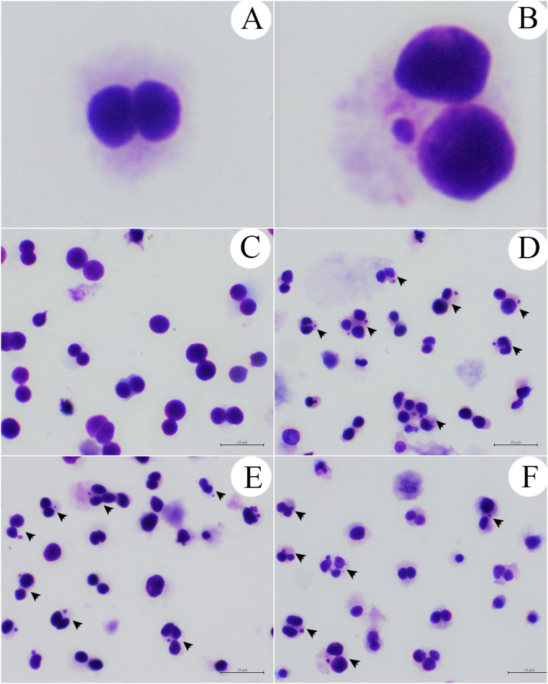
Downloads
References
J Fleurence. Seaweed proteins: Biochemical, nutritional aspects and potential uses. Trends Food Sci. Tech. 1999; 10, 25-8.
ML Wells, P Potin, JS Craigie, JA Raven, SS Merchant, KE Helliwell, AG Smith, ME Camire and SH Brawley. Algae as nutritional and functional food sources: Revisiting our understanding. J. Appl. Phycol. 2017; 29, 949-82.
WB John, RC Brent, HGM Murray, TN Peter and RP Michèle. Marine natural products. Nat. Prod. Rep. 2005; 22, 15-61.
A Jiménez-Escrig, E Gómez-Ordóñez and P Rupérez. Brown and red seaweeds as potential sources of antioxidant nutraceuticals. J. Appl. Phycol. 2012; 24, 1123-32.
TA Fenoradosoa, C Delattre, C Laroche, A Wadouachi, V Dulong, L Picton, P Andriamadio and P Michaud. Highly sulphated galactan from Halymenia durvillei (Halymeniales, Rhodophyta), a red seaweed of Madagascar marine coasts. Int. J. Biol. Macromol. 2009; 45, 140-5.
S Meesala, P Gurung, K Karmodiya, P Subrayan and MG Watve. Isolation and structure elucidation of halymeniaol, a new antimalarial sterol derivative from the red alga Halymenia floresii. J. Asian Nat. Prod. Res. 2018; 20, 391-8.
M Vinosha, S Palanisamy, R Anjali, C Li, K Yelithao, T Marudhupandi, M Tabarsa, SG You and NM Prabhu. Sulfated galactan from Halymenia dilatata enhance the antioxidant properties and prevents Aeromonas hydrophila infection in tilapia fish: In vitro and in vivo study. Int. J. Biol. Macromol. 2020; 158, 569-79.
P Manohong, N Sornkaew, K Meemon, K Chumphoochai, P Sobhon, M Tamtin, J Sichaem, W Mingvanish, C Srisuwannaket, W Mingvanish and N Niamnont. Isolation of 3-(Hydroxyacetyl) indole and Indole-3-carboxylic acid from Red Alga Halymenia durvillei: Their anti-lung cancer cell and in vivo anti-aging activity. Asian J. Chem. 2021; 33, 775-80.
B Halliwell. Antioxidants in human health and disease. Ann. Rev. Nutr. 1996; 16, 33-50.
B Halliwell, JMC Gutteridge and CE Cross. Free radicals, antioxidants and human disease: Where are we now? J. Lab. Clin. Med. 1992; 119, 598-620.
OI Aruoma. Nutrition and health aspects of free radicals and antioxidants. Food Chem. Toxicol. 1994; 32, 671-83.
S Srivastava, S Mishra, J Dewangan, A Divakar, PK Pandey and SK Rath. Principles for in vitro toxicology. In: A Dhawan and S Kwon (Eds.). In vitro toxicology. Academic Press, Massachusetts, 2018, p. 21-43.
AK Jain, D Singh, K Dubey, R Maurya, S Mittal and AK Pandey. Models and methods for in vitro toxicity. In vitro toxicology. Academic Press, Massachusetts, 2018, p. 45-65.
SC Nicklisch and JH Waite. Optimized DPPH assay in a detergent-based buffer system for measuring antioxidant activity of proteins. MethodsX 2014; 1, 233-8.
Z Mellouk, I Benammar, D Krouf, M Goudjil, M Okbi and W Malaisse. Antioxidant properties of the red alga Asparagopsis taxiformis collected on the North West Algerian coast. Exp. Ther. Med. 2017; 13, 32812-90.
A Floegel, DO Kim, SJ Chung, SI Koo and OK Chun. Comparison of ABTS/DPPH assays to measure antioxidant capacity in popular antioxidant-rich US foods. J. Food Compos. Anal. 2011; 24, 1043-8.
V Zacchino, G Centoducati, M Narracci, M Selvaggi and MP Santacroce. Effects of benzo [a] pyrene on gilthead sea bream (Sparus aurata L.) hepatocytes exposed in vitro to short and long term trials. Ital. J. Anim. Sci. 2013; 12, e17.
J Bouayed, L Hoffmann and T Bohn. Total phenolics, flavonoids, anthocyanins and antioxidant activity following simulated gastro-intestinal digestion and dialysis of apple varieties: Bioaccessibility and potential uptake. Food Chem. 2011; 128, 14-21.
LMT Kashani, M Majdzadeh, M Khanavi, M Taghizadeh, N Sadati, N Kahkeshani, M Vatankhah and SN Ostad. Cytotoxic activity of selected Iranian traditional medicinal plants on colon, colorectal and breast cancer cell lines. Arch. Breast. Canc. 2014; 1, 95-8.
K Abe and K Yanagisawa. A new class of rapidly developing mutants in Dictyostelium discoideum: implications for cyclic AMP metabolism and cell differentiation. Dev. Biol. 1983; 95, 200-10.
MF Melzig, G Bader and R Loose. Investigations of the mechanism of membrane activity of selected triterpenoid saponins. Planta Med. 2001; 67, 43-8.
C Gauthier, J Legault, M Piochon-Gauthier and A Pichette. Advances in the synthesis and pharmacological activity of lupane-type triterpenoid saponins. Phytochem. Rev. 2011; 10, 521-44.
A Galanty, M Michalik, Ł Sędek and I Podolak. The influence of LTS-4, a saponoside from Lysimachia thyrsiflora L., on human skin fibroblasts and human melanoma cells. Cell. Mol. Biol. Lett. 2008; 13, 585-98.
P Cmoch, Z Pakulski, J Swaczynová and M Strnad. Synthesis of lupane-type saponins bearing mannosyl and 3, 6-branched trimannosyl residues and their evaluation as anticancer agents. Carbohydr. Res. 2008; 343, 995-1003.
R Tundis, M Bonesi, B Deguin, MR Loizzo, F Menichini, F Conforti, F Tillequin and F Menichini. Cytotoxic activity and inhibitory effect on nitric oxide production of triterpene saponins from the roots of Physospermum verticillatum (Waldst & Kit) (Apiaceae). Bioorg. Med. Chem. 2009; 17, 4542-7.
X Ma, L Xie, L Liu, Q Tang, Z Wan and Y Li. Simultaneous quantification of seven main triterpenoid saponins in Radix et Rhizoma Clematidis by LC-ELSD. Chromatographia 2009; 69, 437-43.
L Mskhiladze, J Legault, S Lavoie, V Mshvildadze, J Kuchukhidze, R Elias and A Pichette. Cytotoxic steroidal saponins from the flowers of Allium leucanthum. Molecules 2008; 13, 2925-34.
A Sánchez-Medina, PC Stevenson, S Habtemariam, LM Peña-Rodríguez, O Corcoran, AI Mallet and NC Veitch. Triterpenoid saponins from a cytotoxic root extract of Sideroxylon foetidissimum subsp. Gaumeri. Phytochemistry 2009; 70, 765-72.
AI Hamed, S Piacente, G Autore, S Marzocco, C Pizza and W Oleszek. Antiproliferative hopane and oleanane glycosides from the roots of Glinus lotoides. Planta Med. 2005; 71, 554-60.
AA Magid, L Voutquenne-Nazabadioko, I Renimel, D Harakat, C Moretti and C Lavaud. Triterpenoid saponins from the stem bark of Caryocar villosum. Phytochemistry 2006; 67, 2096-102.
P Fowler, R Smith, K Smith, J Young, L Jeffrey, P Carmichael, D Kirkland and S Pfuhler. Reduction of misleading (“false”) positive results in mammalian cell genotoxicity assays. III: Sensitivity of human cell types to known genotoxic agents. Mutat. Res. 2014; 767, 28-36.
FM Tonelli, VA Goulart, KN Gomes, MS Ladeira, AK Santos, E Lorençon, LO Ladeira and RR Resende. Graphene-based nanomaterials: Biological and medical applications and toxicity. Nanomedicine 2015; 10, 1-28.
G Gajski, V Garaj-Vrhovac and V Oreščanin. Cytogenetic status and oxidative DNA-damage induced by atorvastatin in human peripheral blood lymphocytes: Standard and Fpg-modified comet assay, Toxicol. Appl. Pharm. 2008; 231, 85-93.
FV Goethem, D Lison and M Kirsch-Volders. Comparative evaluation of the in vitro micronucleus test and the alkaline single cell gel electrophoresis assay for the detection of DNA damaging agents: genotoxic effects of cobalt powder, tungsten carbide and cobalt-tungsten carbide. Mutat. Res. 1997; 392, 31-43.
RF Lee and S Steinert. Use of the single cell gel electrophoresis/comet assay for detecting DNA damage in aquatic (marine and freshwater) animals. Mutat. Res. 2003; 544, 43-64.
M Fenech, M Kirsch-Volders, AT Natarajan, J Surralles, JW Crott, J Parry, H Norppa, DA Eastmond and P Thomas. Molecular mechanisms of micronucleus, nucleoplasmic bridge and nuclear bud formation in mammalian and human cells. Mutagenesis 2011; 26, 125-32.
Downloads
Published
How to Cite
Issue
Section
License

This work is licensed under a Creative Commons Attribution-NonCommercial-NoDerivatives 4.0 International License.


